6FXA

Dimerization domain of TP901-1 CI repressor


Experimental Data Snapshot

  • Method: X-RAY DIFFRACTION
  • Resolution: 1.50 Å
  • R-Value Free: 0.200 
  • R-Value Work: 0.145 
  • R-Value Observed: 0.148 

wwPDB Validation   3D Report Full Report


This is version 1.1 of the entry. See complete history


Literature

Structural basis of the bacteriophage TP901-1 CI repressor dimerization and interaction with DNA.

Rasmussen, K.K.Varming, A.K.Schmidt, S.N.Frandsen, K.E.H.Thulstrup, P.W.Jensen, M.R.Lo Leggio, L.

(2018) FEBS Lett 592: 1738-1750

  • DOI: https://doi.org/10.1002/1873-3468.13060
  • Primary Citation of Related Structures:  
    6FXA

  • PubMed Abstract: 

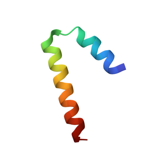
    Temperate bacteriophages are known for their bistability, which in TP901-1 is controlled by two proteins, CI and MOR. Clear 1 repressor (CI) is hexameric and binds three palindromic operator sites via an N-terminal helix-turn-helix domain (NTD). A dimeric form, such as the truncated CI∆58 investigated here, is necessary for high-affinity binding to DNA. The crystal structure of the dimerization region (CTD 1 ) is determined here, showing that it forms a pair of helical hooks. This newly determined structure is used together with the known crystal structure of the CI-NTD and small angle X-ray scattering data, to determine the solution structure of CI∆58 in complex with a palindromic operator site, showing that the two NTDs bind on opposing sides of the DNA helix.


  • Organizational Affiliation

    Department of Chemistry, University of Copenhagen, Denmark.


Macromolecules
Find similar proteins by:  (by identity cutoff)  |  3D Structure
Entity ID: 1
MoleculeChains Sequence LengthOrganismDetailsImage
CI
A, B, C, D, E
A, B, C, D, E, F
35Lactococcus phage TP901-1Mutation(s): 0 
UniProt
Find proteins for O48503 (Lactococcus phage TP901-1)
Explore O48503 
Go to UniProtKB:  O48503
Entity Groups  
Sequence Clusters30% Identity50% Identity70% Identity90% Identity95% Identity100% Identity
UniProt GroupO48503
Sequence Annotations
Expand
  • Reference Sequence
Experimental Data & Validation

Experimental Data

  • Method: X-RAY DIFFRACTION
  • Resolution: 1.50 Å
  • R-Value Free: 0.200 
  • R-Value Work: 0.145 
  • R-Value Observed: 0.148 
  • Space Group: P 21 21 21
Unit Cell:
Length ( Å )Angle ( ˚ )
a = 43.84α = 90
b = 42.56β = 90
c = 101.16γ = 90
Software Package:
Software NamePurpose
PHENIXrefinement
XDSdata reduction
XSCALEdata scaling
PHASERphasing
PDB_EXTRACTdata extraction

Structure Validation

View Full Validation Report


Entry History & Funding Information

Deposition Data


Funding OrganizationLocationGrant Number
Danish Council for Independent ResearchDenmark4002-00107

Revision History  (Full details and data files)

  • Version 1.0: 2018-05-30
    Type: Initial release
  • Version 1.1: 2018-06-13
    Changes: Data collection, Database references