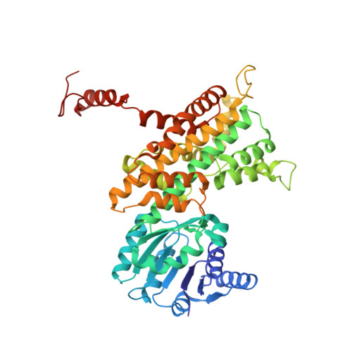
6FQX

Plasmodium falciparum 6-phosphogluconate dehydrogenase in its apo form, in complex with its cofactor NADP+ and in complex with its substrate 6-phosphogluconate


Experimental Data Snapshot

  • Method: X-RAY DIFFRACTION
  • Resolution: 2.80 Å
  • R-Value Free: 0.242 
  • R-Value Work: 0.188 
  • R-Value Observed: 0.189 

wwPDB Validation   3D Report Full Report


This is version 1.3 of the entry. See complete history


Literature

Characterization of Plasmodium falciparum 6-Phosphogluconate Dehydrogenase as an Antimalarial Drug Target.

Haeussler, K.Fritz-Wolf, K.Reichmann, M.Rahlfs, S.Becker, K.

(2018) J Mol Biol 430: 4049-4067

  • DOI: https://doi.org/10.1016/j.jmb.2018.07.030
  • Primary Citation of Related Structures:  
    6FQX, 6FQY, 6FQZ

  • PubMed Abstract: 

    The enzyme 6-phosphogluconate dehydrogenase (6PGD) of the malaria parasite Plasmodium falciparum catalyzes the third step of the pentose phosphate pathway converting 6-phosphogluconate (6PG) to ribulose 5-phosphate. The NADPH produced by 6PGD is crucial for antioxidant defense and redox regulation, and ribose 5-phosphate is essential for DNA and RNA synthesis in the rapidly growing parasite. Thus, 6PGD represents an attractive antimalarial drug target. In this study, we present the X-ray structures of Pf6PGD in native form as well as in complex with 6PG or nicotinamide adenine dinucleotide phosphate (NADP + ) at resolutions of 2.8, 1.9, and 2.9 Å, respectively. The overall structure of the protein is similar to structures of 6PGDs from other species; however, a flexible loop close to the active site rearranges upon binding of 6PG and likely regulates the conformation of the cofactor NADP + . Upon binding of 6PG, the active site loop adopts a closed conformation. In the absence of 6PG, the loop opens and NADP + is bound in a waiting position, indicating that the cofactor and 6PG bind independently from each other. This sequential binding mechanism was supported by kinetic studies on the homodimeric wild-type Pf6PGD. Furthermore, the function of the Plasmodium-specific residue W104L mutant was characterized by site-directed mutagenesis. Notably, the activity of Pf6PGD was found to be post-translationally redox regulated via S-nitrosylation, and screening the Medicines for Malaria Venture Malaria Box identified several compounds with IC 50 s in the low micromolar range. Together with the three-dimensional structure of the protein, this is a promising starting point for further drug discovery approaches.


  • Organizational Affiliation

    Biochemistry and Molecular Biology, Interdisciplinary Research Center, Justus Liebig University, 35392 Giessen, Germany.


Macromolecules
Find similar proteins by:  (by identity cutoff)  |  3D Structure
Entity ID: 1
MoleculeChains Sequence LengthOrganismDetailsImage
6-phosphogluconate dehydrogenase, decarboxylating
A, B, C, D, E
A, B, C, D, E, F, G, H
468Plasmodium falciparum 3D7Mutation(s): 0 
Gene Names: PF14_0520PF3D7_1454700
EC: 1.1.1.44
UniProt
Find proteins for Q8IKT2 (Plasmodium falciparum (isolate 3D7))
Explore Q8IKT2 
Go to UniProtKB:  Q8IKT2
Entity Groups  
Sequence Clusters30% Identity50% Identity70% Identity90% Identity95% Identity100% Identity
UniProt GroupQ8IKT2
Sequence Annotations
Expand
  • Reference Sequence
Small Molecules
Ligands 4 Unique
IDChains Name / Formula / InChI Key2D Diagram3D Interactions
PGE
Query on PGE

Download Ideal Coordinates CCD File 
M [auth A],
S [auth B]
TRIETHYLENE GLYCOL
C6 H14 O4
ZIBGPFATKBEMQZ-UHFFFAOYSA-N
AE3
Query on AE3

Download Ideal Coordinates CCD File 
W [auth D]2-(2-ETHOXYETHOXY)ETHANOL
C6 H14 O3
XXJWXESWEXIICW-UHFFFAOYSA-N
PEG
Query on PEG

Download Ideal Coordinates CCD File 
AA [auth E]
EA [auth F]
GA [auth H]
HA [auth H]
K [auth A]
AA [auth E],
EA [auth F],
GA [auth H],
HA [auth H],
K [auth A],
L [auth A],
R [auth B],
U [auth C],
Z [auth E]
DI(HYDROXYETHYL)ETHER
C4 H10 O3
MTHSVFCYNBDYFN-UHFFFAOYSA-N
GOL
Query on GOL

Download Ideal Coordinates CCD File 
BA [auth F]
CA [auth F]
DA [auth F]
FA [auth H]
I [auth A]
BA [auth F],
CA [auth F],
DA [auth F],
FA [auth H],
I [auth A],
J [auth A],
N [auth B],
O [auth B],
P [auth B],
Q [auth B],
T [auth C],
V [auth D],
X [auth E],
Y [auth E]
GLYCEROL
C3 H8 O3
PEDCQBHIVMGVHV-UHFFFAOYSA-N
Experimental Data & Validation

Experimental Data

  • Method: X-RAY DIFFRACTION
  • Resolution: 2.80 Å
  • R-Value Free: 0.242 
  • R-Value Work: 0.188 
  • R-Value Observed: 0.189 
  • Space Group: P 65
Unit Cell:
Length ( Å )Angle ( ˚ )
a = 134.45α = 90
b = 134.45β = 90
c = 406.05γ = 120
Software Package:
Software NamePurpose
PHENIXrefinement
XDSdata reduction
XSCALEdata scaling
PHENIXphasing

Structure Validation

View Full Validation Report


Entry History & Funding Information

Deposition Data


Funding OrganizationLocationGrant Number
German Research FoundationGermanyBE1540/23-2
Hessian Excellence ProgrammGermanyLoewe center Druid, B3 and E3

Revision History  (Full details and data files)

  • Version 1.0: 2018-08-22
    Type: Initial release
  • Version 1.1: 2018-10-24
    Changes: Data collection, Database references
  • Version 1.2: 2019-01-23
    Changes: Advisory, Data collection, Derived calculations
  • Version 1.3: 2024-01-17
    Changes: Data collection, Database references, Refinement description