


None
Gelin, M., Pochet, S., Labesse, G.To be published.
Experimental Data Snapshot
Starting Model: experimental
View more details
Entity ID: 1 | |||||
|---|---|---|---|---|---|
| Molecule | Chains | Sequence Length | Organism | Details | Image |
| Mitogen-activated protein kinase 1 | 364 | Rattus norvegicus | Mutation(s): 0 Gene Names: Mapk1, Erk2, Mapk, Prkm1 EC: 2.7.11.24 | 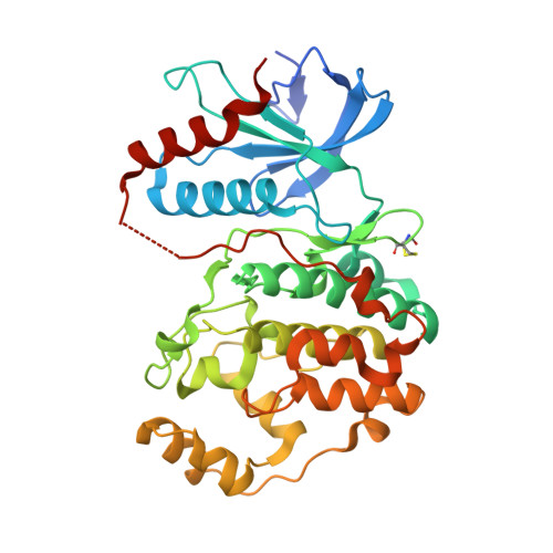 | |
UniProt | |||||
Entity Groups | |||||
| Sequence Clusters | 30% Identity50% Identity70% Identity90% Identity95% Identity100% Identity | ||||
| UniProt Group | P63086 | ||||
Sequence AnnotationsExpand | |||||
Reference Sequence | |||||
| Ligands 3 Unique | |||||
|---|---|---|---|---|---|
| ID | Chains | Name / Formula / InChI Key | 2D Diagram | 3D Interactions | |
| DU8 Download:Ideal Coordinates CCD File | G [auth A] | 3-[6-azanyl-9-[(2~{R},3~{R},4~{S},5~{R})-5-(azidomethyl)-3,4-bis(oxidanyl)oxolan-2-yl]purin-8-yl]sulfanylpropanoic acid C13 H16 N8 O5 S HSUVCMYSEUNGIN-JJNLEZRASA-N |  | ||
| SO4 Download:Ideal Coordinates CCD File | F [auth A] | SULFATE ION O4 S QAOWNCQODCNURD-UHFFFAOYSA-L |  | ||
| DMS Download:Ideal Coordinates CCD File | B [auth A], C [auth A], D [auth A], E [auth A] | DIMETHYL SULFOXIDE C2 H6 O S IAZDPXIOMUYVGZ-UHFFFAOYSA-N |  | ||
| Modified Residues 1 Unique | |||||
|---|---|---|---|---|---|
| ID | Chains | Type | Formula | 2D Diagram | Parent |
| CME Query on CME | A | L-PEPTIDE LINKING | C5 H11 N O3 S2 |  | CYS |
| Length ( Å ) | Angle ( ˚ ) |
|---|---|
| a = 48.66 | α = 90 |
| b = 70.04 | β = 108.62 |
| c = 59.64 | γ = 90 |
| Software Name | Purpose |
|---|---|
| SCALA | data scaling |
| PHENIX | refinement |
| PDB_EXTRACT | data extraction |
| iMOSFLM | data reduction |
| PHENIX | phasing |
| Funding Organization | Location | Grant Number |
|---|---|---|
| FRISBI | France | ANR-10-INSB-05 |


