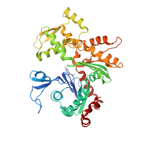
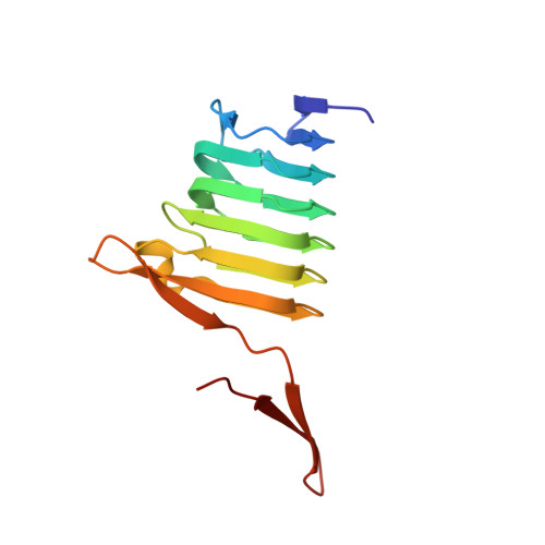
6FM2

CARP domain of mouse cyclase-associated protein 1 (CAP1) bound to ADP-actin


Experimental Data Snapshot

  • Method: X-RAY DIFFRACTION
  • Resolution: 2.80 Å
  • R-Value Free: 0.234 
  • R-Value Work: 0.183 
  • R-Value Observed: 0.186 

Starting Models: experimental
View more details

wwPDB Validation   3D Report Full Report


Ligand Structure Quality Assessment 


This is version 1.3 of the entry. See complete history


Literature

Structural basis of actin monomer re-charging by cyclase-associated protein.

Kotila, T.Kogan, K.Enkavi, G.Guo, S.Vattulainen, I.Goode, B.L.Lappalainen, P.

(2018) Nat Commun 9: 1892-1892

  • DOI: https://doi.org/10.1038/s41467-018-04231-7
  • Primary Citation of Related Structures:  
    6FM2

  • PubMed Abstract: 

    Actin polymerization powers key cellular processes, including motility, morphogenesis, and endocytosis. The actin turnover cycle depends critically on "re-charging" of ADP-actin monomers with ATP, but whether this reaction requires dedicated proteins in cells, and the underlying mechanism, have remained elusive. Here we report that nucleotide exchange catalyzed by the ubiquitous cytoskeletal regulator cyclase-associated protein (CAP) is critical for actin-based processes in vivo. We determine the structure of the CAP-actin complex, which reveals that nucleotide exchange occurs in a compact, sandwich-like complex formed between the dimeric actin-binding domain of CAP and two ADP-actin monomers. In the crystal structure, the C-terminal tail of CAP associates with the nucleotide-sensing region of actin, and this interaction is required for rapid re-charging of actin by both yeast and mammalian CAPs. These data uncover the conserved structural basis and biological role of protein-catalyzed re-charging of actin monomers.


  • Organizational Affiliation

    Institute of Biotechnology, University of Helsinki, 00014, Helsinki, Finland.


Macromolecules
Find similar proteins by:  (by identity cutoff)  |  3D Structure
Entity ID: 1
MoleculeChains Sequence LengthOrganismDetailsImage
Actin, alpha skeletal muscle375Oryctolagus cuniculusMutation(s): 0 
UniProt
Find proteins for P68135 (Oryctolagus cuniculus)
Explore P68135 
Go to UniProtKB:  P68135
Entity Groups  
Sequence Clusters30% Identity50% Identity70% Identity90% Identity95% Identity100% Identity
UniProt GroupP68135
Sequence Annotations
Expand
  • Reference Sequence
Find similar proteins by:  (by identity cutoff)  |  3D Structure
Entity ID: 2
MoleculeChains Sequence LengthOrganismDetailsImage
Adenylyl cyclase-associated protein 1158Mus musculusMutation(s): 0 
Gene Names: Cap1Cap
UniProt
Find proteins for P40124 (Mus musculus)
Explore P40124 
Go to UniProtKB:  P40124
Entity Groups  
Sequence Clusters30% Identity50% Identity70% Identity90% Identity95% Identity100% Identity
UniProt GroupP40124
Sequence Annotations
Expand
  • Reference Sequence
Small Molecules
Modified Residues  1 Unique
IDChains TypeFormula2D DiagramParent
HIC
Query on HIC
A
L-PEPTIDE LINKINGC7 H11 N3 O2HIS
Experimental Data & Validation

Experimental Data

  • Method: X-RAY DIFFRACTION
  • Resolution: 2.80 Å
  • R-Value Free: 0.234 
  • R-Value Work: 0.183 
  • R-Value Observed: 0.186 
  • Space Group: P 61 2 2
Unit Cell:
Length ( Å )Angle ( ˚ )
a = 73.833α = 90
b = 73.833β = 90
c = 453.377γ = 120
Software Package:
Software NamePurpose
BUSTERrefinement
XDSdata reduction
Aimlessdata scaling
BALBESphasing
Cootmodel building

Structure Validation

View Full Validation Report


Ligand Structure Quality Assessment 


Entry History & Funding Information

Deposition Data


Funding OrganizationLocationGrant Number
Academy of FinlandFinland272130
Academy of FinlandFinland307415
European Research CouncilFinland290974
National Institutes of Health/National Human Genome Research Institute (NIH/NHGRI)United StatesR01 GM063691
National Science Foundation (United States)United StatesDMR-1420382

Revision History  (Full details and data files)

  • Version 1.0: 2018-05-16
    Type: Initial release
  • Version 1.1: 2018-05-23
    Changes: Data collection, Database references
  • Version 1.2: 2022-03-30
    Changes: Author supporting evidence, Database references, Derived calculations
  • Version 1.3: 2024-01-17
    Changes: Data collection, Refinement description