6F9G | pdb_00006f9g

Ligand binding domain of P. putida KT2440 polyamine chemorecpetors McpU in complex putrescine.


Experimental Data Snapshot

  • Method: X-RAY DIFFRACTION
  • Resolution: 2.39 Å
  • R-Value Free: 
    0.234 (Depositor), 0.234 (DCC) 
  • R-Value Work: 
    0.195 (Depositor), 0.196 (DCC) 
  • R-Value Observed: 
    0.197 (Depositor) 

wwPDB Validation 3D Report Full Report

Validation slider image for 6F9G

Ligand Structure Quality Assessment 


This is version 1.3 of the entry. See complete history

Literature

Structural Basis for Polyamine Binding at the dCACHE Domain of the McpU Chemoreceptor from Pseudomonas putida.

Gavira, J.A.Ortega, A.Martin-Mora, D.Conejero-Muriel, M.T.Corral-Lugo, A.Morel, B.Matilla, M.A.Krell, T.

(2018) J Mol Biology 430: 1950-1963

  • DOI: https://doi.org/10.1016/j.jmb.2018.05.008
  • Primary Citation Related Structures: 
    6F9G

  • PubMed Abstract: 

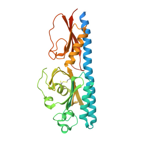
    Many bacteria can move chemotactically to a variety of compounds and the recognition of chemoeffectors by the chemoreceptor ligand binding domain (LBD) defines the specificity of response. Many chemoreceptors were found to recognize different amino and organic acids, but the McpU chemoreceptor from Pseudomonas putida was identified as the first chemoreceptor that bound specifically polyamines. We report here the three-dimensional structure of McpU-LBD in complex with putrescine at a resolution of 2.4 Å, which fitted well a solution structure generated by small-angle X-ray scattering. Putrescine bound to a negatively charged pocket in the membrane distal module of McpU-LBD. Similarities exist in the binding of putrescine to McpU-LBD and taurine to the LBD of the Mlp37 chemoreceptor of Vibrio cholerae. In both structures, the primary amino group of the respective ligand is recognized by hydrogen bonds established by two aspartate and a tyrosine side chain. This feature may be used to predict the ligands of chemoreceptors with unknown function. Analytical ultracentrifugation revealed that McpU-LBD is monomeric in solution and that ligand binding does not alter this oligomeric state. This sensing mode thus differs from that of the well-characterised four-helix bundle domains where ligands bind to two sites at the LBD dimer interface. Although there appear to be different sensing modes, results are discussed in the context of data, indicating that chemoreceptors employ the same mechanism of transmembrane signaling. This work enhances our understanding of CACHE domains, which are the most abundant sensor domains in bacterial chemoreceptors and sensor kinases.


  • Organizational Affiliation
    • Laboratory of Crystallographic Studies, IACT, (CSIC-UGR), Avenida de las Palmeras 4, 18100 Armilla, Granada, Spain.

Macromolecule Content 

  • Total Structure Weight: 174.68 kDa 
  • Atom Count: 11,034 
  • Modeled Residue Count: 1,305 
  • Deposited Residue Count: 1,605 
  • Unique protein chains: 1

Macromolecules

Find similar proteins by:|  3D Structure
Entity ID: 1
MoleculeChains  Sequence LengthOrganismDetailsImage
Methyl-accepting chemotaxis protein McpU
A, B, C, D, E
321Pseudomonas putida KT2440Mutation(s): 0 
Gene Names: mcpUPP_1228
UniProt
Find proteins for Q88NI1 (Pseudomonas putida (strain ATCC 47054 / DSM 6125 / CFBP 8728 / NCIMB 11950 / KT2440))
Explore Q88NI1 
Go to UniProtKB:  Q88NI1
Entity Groups
Sequence Clusters30% Identity50% Identity70% Identity90% Identity95% Identity100% Identity
UniProt GroupQ88NI1
Sequence Annotations
Expand
Reference Sequence

Small Molecules

Ligands 3 Unique
IDChains Name / Formula / InChI Key2D Diagram3D Interactions
GOL

Query on GOL


Download:Ideal Coordinates CCD File
G [auth A],
M [auth D],
N [auth D],
P [auth E],
Q [auth E]
GLYCEROL
C3 H8 O3
PEDCQBHIVMGVHV-UHFFFAOYSA-N
PUT
(Subject of Investigation/LOI)

Query on PUT


Download:Ideal Coordinates CCD File
F [auth A],
H [auth B],
I [auth C],
K [auth D],
O [auth E]
1,4-DIAMINOBUTANE
C4 H12 N2
KIDHWZJUCRJVML-UHFFFAOYSA-N
ACT

Query on ACT


Download:Ideal Coordinates CCD File
J [auth C],
L [auth D]
ACETATE ION
C2 H3 O2
QTBSBXVTEAMEQO-UHFFFAOYSA-M

Experimental Data & Validation

Experimental Data

  • Method: X-RAY DIFFRACTION
  • Resolution: 2.39 Å
  • R-Value Free:  0.234 (Depositor), 0.234 (DCC) 
  • R-Value Work:  0.195 (Depositor), 0.196 (DCC) 
  • R-Value Observed: 0.197 (Depositor) 
Space Group: C 2 2 21
Unit Cell:
Length ( Å )Angle ( ˚ )
a = 133.984α = 90
b = 164.521β = 90
c = 123.612γ = 90
Software Package:
Software NamePurpose
PHENIXrefinement
XDSdata reduction
Aimlessdata scaling
Auto-Rickshawphasing

Structure Validation

View Full Validation Report


Ligand Structure Quality Assessment 


Entry History 

& Funding Information

Deposition Data


Funding OrganizationLocationGrant Number
MICINNSpainBIO2013-4297-P

Revision History  (Full details and data files)

  • Version 1.0: 2018-03-28
    Type: Initial release
  • Version 1.1: 2018-05-23
    Changes: Data collection, Database references, Source and taxonomy
  • Version 1.2: 2018-06-13
    Changes: Data collection, Database references
  • Version 1.3: 2024-10-16
    Changes: Data collection, Database references, Structure summary