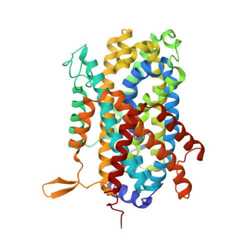
6F34

Crystal structure of a bacterial cationic amino acid transporter (CAT) homologue bound to Arginine.


Experimental Data Snapshot

  • Method: X-RAY DIFFRACTION
  • Resolution: 3.13 Å
  • R-Value Free: 0.262 
  • R-Value Work: 0.231 
  • R-Value Observed: 0.233 

Starting Model: experimental
View more details

wwPDB Validation   3D Report Full Report


Ligand Structure Quality Assessment 


This is version 1.2 of the entry. See complete history


Literature

Structural basis for amino acid transport by the CAT family of SLC7 transporters.

Jungnickel, K.E.J.Parker, J.L.Newstead, S.

(2018) Nat Commun 9: 550-550

  • DOI: https://doi.org/10.1038/s41467-018-03066-6
  • Primary Citation of Related Structures:  
    5OQT, 6F34

  • PubMed Abstract: 

    Amino acids play essential roles in cell biology as regulators of metabolic pathways. Arginine in particular is a major signalling molecule inside the cell, being a precursor for both l-ornithine and nitric oxide (NO) synthesis and a key regulator of the mTORC1 pathway. In mammals, cellular arginine availability is determined by members of the solute carrier (SLC) 7 family of cationic amino acid transporters. Whereas CAT-1 functions to supply cationic amino acids for cellular metabolism, CAT-2A and -2B are required for macrophage activation and play important roles in regulating inflammation. Here, we present the crystal structure of a close homologue of the mammalian CAT transporters that reveals how these proteins specifically recognise arginine. Our structural and functional data provide a model for cationic amino acid transport in mammalian cells and reveals mechanistic insights into proton-coupled, sodium-independent amino acid transport in the wider APC superfamily.


  • Organizational Affiliation

    Department of Biochemistry, University of Oxford, South Parks Road, Oxford, OX1 3QU, UK.


Macromolecules
Find similar proteins by:  (by identity cutoff)  |  3D Structure
Entity ID: 1
MoleculeChains Sequence LengthOrganismDetailsImage
Amino acid transporter461Geobacillus kaustophilus HTA426Mutation(s): 0 
Gene Names: GK0930ApcT
Membrane Entity: Yes 
UniProt
Find proteins for Q5L1G5 (Geobacillus kaustophilus (strain HTA426))
Explore Q5L1G5 
Go to UniProtKB:  Q5L1G5
Entity Groups  
Sequence Clusters30% Identity50% Identity70% Identity90% Identity95% Identity100% Identity
UniProt GroupQ5L1G5
Sequence Annotations
Expand
  • Reference Sequence
Find similar proteins by:  (by identity cutoff)  |  3D Structure
Entity ID: 2
MoleculeChains Sequence LengthOrganismDetailsImage
MgtSB [auth C]27Escherichia coli K-12Mutation(s): 0 
Gene Names: yneMb4599JW1527.1
Membrane Entity: Yes 
UniProt
Find proteins for A5A616 (Escherichia coli (strain K12))
Explore A5A616 
Go to UniProtKB:  A5A616
Entity Groups  
Sequence Clusters30% Identity50% Identity70% Identity90% Identity95% Identity100% Identity
UniProt GroupA5A616
Sequence Annotations
Expand
  • Reference Sequence
Experimental Data & Validation

Experimental Data

  • Method: X-RAY DIFFRACTION
  • Resolution: 3.13 Å
  • R-Value Free: 0.262 
  • R-Value Work: 0.231 
  • R-Value Observed: 0.233 
  • Space Group: P 21 21 21
Unit Cell:
Length ( Å )Angle ( ˚ )
a = 77.037α = 90
b = 82.699β = 90
c = 118.782γ = 90
Software Package:
Software NamePurpose
Aimlessdata scaling
PHENIXrefinement
PDB_EXTRACTdata extraction
DIALSdata reduction
PHASERphasing

Structure Validation

View Full Validation Report


Ligand Structure Quality Assessment 


Entry History & Funding Information

Deposition Data


Funding OrganizationLocationGrant Number
Wellcome TrustUnited Kingdom102890/Z/13/Z

Revision History  (Full details and data files)

  • Version 1.0: 2018-02-14
    Type: Initial release
  • Version 1.1: 2018-02-21
    Changes: Database references
  • Version 1.2: 2024-01-17
    Changes: Data collection, Database references, Refinement description