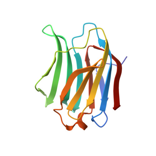
Elucidation of Hydrogen Bonding Patterns in Ligand-Free, Lactose- and Glycerol-Bound Galectin-3C by Neutron Crystallography to Guide Drug Design.
Manzoni, F., Wallerstein, J., Schrader, T.E., Ostermann, A., Coates, L., Akke, M., Blakeley, M.P., Oksanen, E., Logan, D.T.(2018) J Med Chem 61: 4412-4420
- PubMed: 29672051
- DOI: https://doi.org/10.1021/acs.jmedchem.8b00081
- Primary Citation of Related Structures:
6EXY, 6EYM, 6F2Q - PubMed Abstract:
The medically important drug target galectin-3 binds galactose-containing moieties on glycoproteins through an intricate pattern of hydrogen bonds to a largely polar surface-exposed binding site. All successful inhibitors of galectin-3 to date have been based on mono- or disaccharide cores closely resembling natural ligands. A detailed understanding of the H-bonding networks in these natural ligands will provide an improved foundation for the design of novel inhibitors. Neutron crystallography is an ideal technique to reveal the geometry of hydrogen bonds because the positions of hydrogen atoms are directly detected rather than being inferred from the positions of heavier atoms as in X-ray crystallography. We present three neutron crystal structures of the C-terminal carbohydrate recognition domain of galectin-3: the ligand-free form and the complexes with the natural substrate lactose and with glycerol, which mimics important interactions made by lactose. The neutron crystal structures reveal unambiguously the exquisite fine-tuning of the hydrogen bonding pattern in the binding site to the natural disaccharide ligand. The ligand-free structure shows that most of these hydrogen bonds are preserved even when the polar groups of the ligand are replaced by water molecules. The protonation states of all histidine residues in the protein are also revealed and correlate well with NMR observations. The structures give a solid starting point for molecular dynamics simulations and computational estimates of ligand binding affinity that will inform future drug design.
- Department of Biochemistry & Structural Biology , Lund University , S-221 00 Lund , Sweden.
Organizational Affiliation:


