6F29 | pdb_00006f29

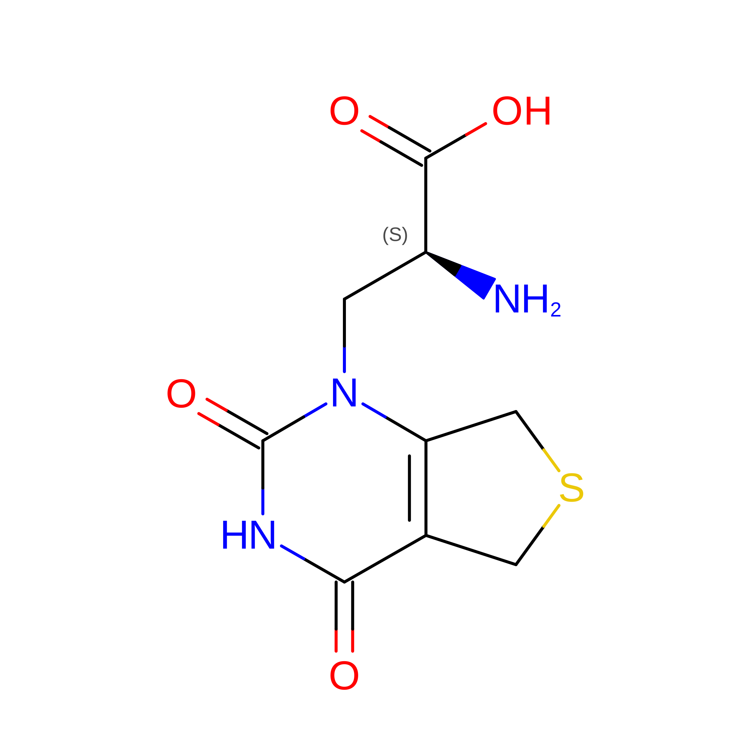
Crystal structure of the kainate receptor GluK3 ligand binding domain in complex with (S)-1-[2-Amino-2-carboxyethyl]-5,7-dihydrothieno[3,4-d]pyrimidin-2,4(1H,3H)-dione at resolution 2.6A


Experimental Data Snapshot

  • Method: X-RAY DIFFRACTION
  • Resolution: 2.60 Å
  • R-Value Free: 
    0.248 (Depositor), 0.246 (DCC) 
  • R-Value Work: 
    0.192 (Depositor), 0.191 (DCC) 
  • R-Value Observed: 
    0.195 (Depositor) 

Starting Model: experimental
View more details

wwPDB Validation 3D Report Full Report

Validation slider image for 6F29

Ligand Structure Quality Assessment 


This is version 1.4 of the entry. See complete history. 

Literature

( S)-2-Amino-3-(5-methyl-3-hydroxyisoxazol-4-yl)propanoic Acid (AMPA) and Kainate Receptor Ligands: Further Exploration of Bioisosteric Replacements and Structural and Biological Investigation.

Brogi, S., Brindisi, M., Butini, S., Kshirsagar, G.U., Maramai, S., Chemi, G., Gemma, S., Campiani, G., Novellino, E., Fiorenzani, P., Pinassi, J., Aloisi, A.M., Gynther, M., Venskutonyte, R., Han, L., Frydenvang, K., Kastrup, J.S., Pickering, D.S.

(2018) J Med Chem 61: 2124-2130

  • DOI: https://doi.org/10.1021/acs.jmedchem.8b00099
  • Primary Citation Related Structures: 
    6F28, 6F29

  • PubMed Abstract: 

    Starting from 1-4 and 7 structural templates, analogues based on bioisosteric replacements (5a-c vs 1, 2 and 6 vs 7) were synthesized for completing the SAR analysis. Interesting binding properties at GluA2, GluK1, and GluK3 receptors were discovered. The requirements for GluK3 interaction were elucidated by determining the X-ray structures of the GluK3-LBD with 2 and 5c and by computational studies. Antinociceptive potential was demonstrated for GluK1 partial agonist 3 and antagonist 7 (2 mg/kg ip).


  • Organizational Affiliation: 
    • Department of Biotechnology, Chemistry and Pharmacy, (DoE 2018-2022) NatSynDrugs , University of Siena , Via A. Moro 2 , 53100 Siena , Italy.

Macromolecule Content 

  • Total Structure Weight: 29.53 kDa 
  • Atom Count: 2,088 
  • Modeled Residue Count: 254 
  • Deposited Residue Count: 258 
  • Unique protein chains: 1

Macromolecules

Find similar proteins by:|  3D Structure
Entity ID: 1
MoleculeChains  Sequence LengthOrganismDetailsImage
Glutamate receptor ionotropic, kainate 3,Glutamate receptor ionotropic, kainate 3258Rattus norvegicusMutation(s): 0 
Gene Names: Grik3, Glur7
UniProt
Find proteins for P42264 (Rattus norvegicus)
Explore P42264 
Go to UniProtKB:  P42264
Entity Groups
Sequence Clusters30% Identity50% Identity70% Identity90% Identity95% Identity100% Identity
UniProt GroupP42264
Sequence Annotations
Expand
Reference Sequence

Small Molecules

Experimental Data & Validation

Experimental Data

  • Method: X-RAY DIFFRACTION
  • Resolution: 2.60 Å
  • R-Value Free:  0.248 (Depositor), 0.246 (DCC) 
  • R-Value Work:  0.192 (Depositor), 0.191 (DCC) 
  • R-Value Observed: 0.195 (Depositor) 
Space Group: P 41 2 2
Unit Cell:
Length ( Å )Angle ( ˚ )
a = 68.04α = 90
b = 68.04β = 90
c = 126.86γ = 90
Software Package:
Software NamePurpose
SCALAdata scaling
PHENIXrefinement
PDB_EXTRACTdata extraction
iMOSFLMdata reduction
PHASERphasing

Structure Validation

View Full Validation Report


Ligand Structure Quality Assessment 


Entry History 

Deposition Data

Revision History  (Full details and data files)

  • Version 1.0: 2018-02-28
    Type: Initial release
  • Version 1.1: 2018-03-07
    Changes: Database references
  • Version 1.2: 2018-03-21
    Changes: Database references
  • Version 1.3: 2024-01-17
    Changes: Data collection, Database references, Derived calculations, Refinement description
  • Version 1.4: 2024-11-13
    Changes: Structure summary