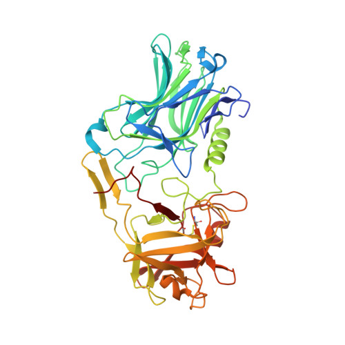
6F0P

Botulinum neurotoxin A4 Hc domain


Experimental Data Snapshot

  • Method: X-RAY DIFFRACTION
  • Resolution: 1.34 Å
  • R-Value Free: 0.158 
  • R-Value Work: 0.144 
  • R-Value Observed: 0.144 

Starting Model: experimental
View more details

wwPDB Validation   3D Report Full Report


This is version 1.3 of the entry. See complete history


Literature

High resolution crystal structures of Clostridium botulinum neurotoxin A3 and A4 binding domains.

Davies, J.R.Rees, J.Liu, S.M.Acharya, K.R.

(2018) J Struct Biol 202: 113-117

  • DOI: https://doi.org/10.1016/j.jsb.2017.12.010
  • Primary Citation of Related Structures:  
    6F0O, 6F0P

  • PubMed Abstract: 

    Clostridium botulinum neurotoxins (BoNTs) cause the life-threatening condition, botulism. However, while they have the potential to cause serious harm, they are increasingly being utilised for therapeutic applications. BoNTs comprise of seven distinct serotypes termed BoNT/A through BoNT/G, with the most widely characterised being sub-serotype BoNT/A1. Each BoNT consists of three structurally distinct domains, a binding domain (H C ), a translocation domain (H N ), and a proteolytic domain (LC). The H C domain is responsible for the highly specific targeting of the neurotoxin to neuronal cell membranes. Here, we present two high-resolution structures of the binding domain of subtype BoNT/A3 (H C /A3) and BoNT/A4 (H C /A4) at 1.6 Å and 1.34 Å resolution, respectively. The structures of both proteins share a high degree of similarity to other known BoNT H C domains whilst containing some subtle differences, and are of benefit to research into therapeutic neurotoxins with novel characteristics.


  • Organizational Affiliation

    Department of Biology and Biochemistry, Claverton Down, University of Bath, Bath BA2 7AY, UK.


Macromolecules
Find similar proteins by:  (by identity cutoff)  |  3D Structure
Entity ID: 1
MoleculeChains Sequence LengthOrganismDetailsImage
Neurotoxin type A433Clostridium botulinumMutation(s): 0 
UniProt
Find proteins for Q3LRX8 (Clostridium botulinum)
Explore Q3LRX8 
Go to UniProtKB:  Q3LRX8
Entity Groups  
Sequence Clusters30% Identity50% Identity70% Identity90% Identity95% Identity100% Identity
UniProt GroupQ3LRX8
Sequence Annotations
Expand
  • Reference Sequence
Small Molecules
Ligands 4 Unique
IDChains Name / Formula / InChI Key2D Diagram3D Interactions
BTB
Query on BTB

Download Ideal Coordinates CCD File 
J [auth A]2-[BIS-(2-HYDROXY-ETHYL)-AMINO]-2-HYDROXYMETHYL-PROPANE-1,3-DIOL
C8 H19 N O5
OWMVSZAMULFTJU-UHFFFAOYSA-N
PEG
Query on PEG

Download Ideal Coordinates CCD File 
C [auth A],
D [auth A]
DI(HYDROXYETHYL)ETHER
C4 H10 O3
MTHSVFCYNBDYFN-UHFFFAOYSA-N
EDO
Query on EDO

Download Ideal Coordinates CCD File 
E [auth A],
F [auth A],
G [auth A],
H [auth A],
I [auth A]
1,2-ETHANEDIOL
C2 H6 O2
LYCAIKOWRPUZTN-UHFFFAOYSA-N
NI
Query on NI

Download Ideal Coordinates CCD File 
B [auth A]NICKEL (II) ION
Ni
VEQPNABPJHWNSG-UHFFFAOYSA-N
Experimental Data & Validation

Experimental Data

  • Method: X-RAY DIFFRACTION
  • Resolution: 1.34 Å
  • R-Value Free: 0.158 
  • R-Value Work: 0.144 
  • R-Value Observed: 0.144 
  • Space Group: P 21 3
Unit Cell:
Length ( Å )Angle ( ˚ )
a = 115.888α = 90
b = 115.888β = 90
c = 115.888γ = 90
Software Package:
Software NamePurpose
REFMACrefinement
DIALSdata reduction
Aimlessdata scaling
PHASERphasing

Structure Validation

View Full Validation Report


Entry History 

Deposition Data

Revision History  (Full details and data files)

  • Version 1.0: 2018-01-10
    Type: Initial release
  • Version 1.1: 2018-04-04
    Changes: Data collection, Database references
  • Version 1.2: 2024-01-17
    Changes: Data collection, Database references, Refinement description
  • Version 1.3: 2024-11-06
    Changes: Structure summary