6F0N

GLIC mutant E82A


Experimental Data Snapshot

  • Method: X-RAY DIFFRACTION
  • Resolution: 3.20 Å
  • R-Value Free: 0.233 
  • R-Value Work: 0.209 
  • R-Value Observed: 0.211 

wwPDB Validation   3D Report Full Report


Ligand Structure Quality Assessment 


This is version 1.0 of the entry. See complete history


Literature

Full mutational mapping of titratable residues helps to identify proton-sensors involved in the control of channel gating in the Gloeobacter violaceus pentameric ligand-gated ion channel.

Nemecz, A.Hu, H.Fourati, Z.Van Renterghem, C.Delarue, M.Corringer, P.J.

(2017) PLoS Biol 15: e2004470-e2004470

  • DOI: https://doi.org/10.1371/journal.pbio.2004470
  • Primary Citation of Related Structures:  
    6F0I, 6F0J, 6F0M, 6F0N, 6F0R, 6F0U, 6F0V, 6F0Z, 6F10, 6F11, 6F12, 6F13, 6F15, 6F16

  • PubMed Abstract: 

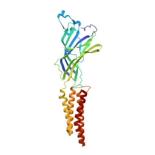
    The Gloeobacter violaceus ligand-gated ion channel (GLIC) has been extensively studied by X-ray crystallography and other biophysical techniques. This provided key insights into the general gating mechanism of pentameric ligand-gated ion channel (pLGIC) signal transduction. However, the GLIC is activated by lowering the pH and the location of its putative proton activation site(s) still remain(s) unknown. To this end, every Asp, Glu, and His residue was mutated individually or in combination and investigated by electrophysiology. In addition to the mutational analysis, key mutations were structurally resolved to address whether particular residues contribute to proton sensing, or alternatively to GLIC-gating, independently of the side chain protonation. The data show that multiple residues located below the orthosteric site, notably E26, D32, E35, and D122 in the lower part of the extracellular domain (ECD), along with E222, H235, E243, and H277 in the transmembrane domain (TMD), alter GLIC activation. D122 and H235 were found to also alter GLIC expression. E35 is identified as a key proton-sensing residue, whereby neutralization of its side chain carboxylate stabilizes the active state. Thus, proton activation occurs allosterically to the orthosteric site, at the level of multiple loci with a key contribution of the coupling interface between the ECD and TMD.


  • Organizational Affiliation

    Unité Récepteurs-Canaux, Unité Mixte de Recherche 3571 du Centre National de la Recherche Scientifique, Institut Pasteur, Paris, France.


Macromolecules
Find similar proteins by:  (by identity cutoff)  |  3D Structure
Entity ID: 1
MoleculeChains Sequence LengthOrganismDetailsImage
Proton-gated ion channel
A, B, C, D, E
317Gloeobacter violaceusMutation(s): 1 
Gene Names: glvIglr4197
Membrane Entity: Yes 
UniProt
Find proteins for Q7NDN8 (Gloeobacter violaceus (strain ATCC 29082 / PCC 7421))
Explore Q7NDN8 
Go to UniProtKB:  Q7NDN8
Entity Groups  
Sequence Clusters30% Identity50% Identity70% Identity90% Identity95% Identity100% Identity
UniProt GroupQ7NDN8
Sequence Annotations
Expand
  • Reference Sequence
Small Molecules
Ligands 5 Unique
IDChains Name / Formula / InChI Key2D Diagram3D Interactions
PLC
Query on PLC

Download Ideal Coordinates CCD File 
CA [auth D],
G [auth A],
IA [auth E],
P [auth B],
V [auth C]
DIUNDECYL PHOSPHATIDYL CHOLINE
C32 H65 N O8 P
IJFVSSZAOYLHEE-SSEXGKCCSA-O
LMT
Query on LMT

Download Ideal Coordinates CCD File 
GA [auth D]
M [auth A]
MA [auth E]
N [auth A]
T [auth B]
GA [auth D],
M [auth A],
MA [auth E],
N [auth A],
T [auth B],
Z [auth C]
DODECYL-BETA-D-MALTOSIDE
C24 H46 O11
NLEBIOOXCVAHBD-QKMCSOCLSA-N
ACT
Query on ACT

Download Ideal Coordinates CCD File 
BA [auth D]
F [auth A]
FA [auth D]
HA [auth E]
L [auth A]
BA [auth D],
F [auth A],
FA [auth D],
HA [auth E],
L [auth A],
LA [auth E],
O [auth B],
S [auth B],
U [auth B],
Y [auth C]
ACETATE ION
C2 H3 O2
QTBSBXVTEAMEQO-UHFFFAOYSA-M
CL
Query on CL

Download Ideal Coordinates CCD File 
DA [auth D]
H [auth A]
I [auth A]
J [auth A]
JA [auth E]
DA [auth D],
H [auth A],
I [auth A],
J [auth A],
JA [auth E],
Q [auth B],
W [auth C]
CHLORIDE ION
Cl
VEXZGXHMUGYJMC-UHFFFAOYSA-M
NA
Query on NA

Download Ideal Coordinates CCD File 
AA [auth D]
EA [auth D]
K [auth A]
KA [auth E]
R [auth B]
AA [auth D],
EA [auth D],
K [auth A],
KA [auth E],
R [auth B],
X [auth C]
SODIUM ION
Na
FKNQFGJONOIPTF-UHFFFAOYSA-N
Experimental Data & Validation

Experimental Data

  • Method: X-RAY DIFFRACTION
  • Resolution: 3.20 Å
  • R-Value Free: 0.233 
  • R-Value Work: 0.209 
  • R-Value Observed: 0.211 
  • Space Group: C 1 2 1
Unit Cell:
Length ( Å )Angle ( ˚ )
a = 180.22α = 90
b = 132.6β = 101.65
c = 159.03γ = 90
Software Package:
Software NamePurpose
BUSTERrefinement
XDSdata reduction
Aimlessdata scaling
PHASERphasing

Structure Validation

View Full Validation Report


Ligand Structure Quality Assessment 


Entry History 

Deposition Data

Revision History  (Full details and data files)

  • Version 1.0: 2018-01-10
    Type: Initial release