6EWP

Mus musculus CEP120 third C2 domain (C2C)


Experimental Data Snapshot

  • Method: X-RAY DIFFRACTION
  • Resolution: 1.85 Å
  • R-Value Free: 0.227 
  • R-Value Work: 0.207 
  • R-Value Observed: 0.208 

wwPDB Validation   3D Report Full Report


This is version 1.1 of the entry. See complete history


Literature

Disease-Associated Mutations in CEP120 Destabilize the Protein and Impair Ciliogenesis.

Joseph, N.Al-Jassar, C.Johnson, C.M.Andreeva, A.Barnabas, D.D.Freund, S.M.V.Gergely, F.van Breugel, M.

(2018) Cell Rep 23: 2805-2818

  • DOI: https://doi.org/10.1016/j.celrep.2018.04.100
  • Primary Citation of Related Structures:  
    6EWG, 6EWH, 6EWI, 6EWL, 6EWP

  • PubMed Abstract: 

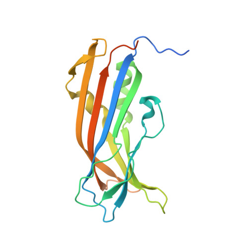
    Ciliopathies are a group of genetic disorders caused by a failure to form functional cilia. Due to a lack of structural information, it is currently poorly understood how ciliopathic mutations affect protein functionality to give rise to the underlying disease. Using X-ray crystallography, we show that the ciliopathy-associated centriolar protein CEP120 contains three C2 domains. The point mutations V194A and A199P, which cause Joubert syndrome (JS) and Jeune asphyxiating thoracic dystrophy (JATD), respectively, both reduce the thermostability of the second C2 domain by targeting residues that point toward its hydrophobic core. Genome-engineered cells homozygous for these mutations have largely normal centriole numbers but show reduced CEP120 levels, compromised recruitment of distal centriole markers, and deficient cilia formation. Our results provide insight into the disease mechanism of two ciliopathic mutations in CEP120, identify putative binding partners of CEP120 C2B, and suggest a complex genotype-phenotype relation of the CEP120 ciliopathy alleles.


  • Organizational Affiliation

    Cancer Research UK Cambridge Institute, University of Cambridge, Li Ka Shing Centre, Robinson Way, Cambridge CB2 0RE, UK.


Macromolecules
Find similar proteins by:  (by identity cutoff)  |  3D Structure
Entity ID: 1
MoleculeChains Sequence LengthOrganismDetailsImage
Centrosomal protein of 120 kDa
A, B, C
201Mus musculusMutation(s): 0 
Gene Names: Cep120Ccdc100
UniProt
Find proteins for Q7TSG1 (Mus musculus)
Explore Q7TSG1 
Go to UniProtKB:  Q7TSG1
Entity Groups  
Sequence Clusters30% Identity50% Identity70% Identity90% Identity95% Identity100% Identity
UniProt GroupQ7TSG1
Sequence Annotations
Expand
  • Reference Sequence
Small Molecules
Ligands 1 Unique
IDChains Name / Formula / InChI Key2D Diagram3D Interactions
P6G
Query on P6G

Download Ideal Coordinates CCD File 
D [auth A]HEXAETHYLENE GLYCOL
C12 H26 O7
IIRDTKBZINWQAW-UHFFFAOYSA-N
Experimental Data & Validation

Experimental Data

  • Method: X-RAY DIFFRACTION
  • Resolution: 1.85 Å
  • R-Value Free: 0.227 
  • R-Value Work: 0.207 
  • R-Value Observed: 0.208 
  • Space Group: P 61
Unit Cell:
Length ( Å )Angle ( ˚ )
a = 107.008α = 90
b = 107.008β = 90
c = 141.762γ = 120
Software Package:
Software NamePurpose
REFMACrefinement
MOSFLMdata reduction
Aimlessdata scaling
SHELXphasing

Structure Validation

View Full Validation Report


Entry History & Funding Information

Deposition Data


Funding OrganizationLocationGrant Number
Medical Research Council (United Kingdom)United KingdomMC_UP_1201/3

Revision History  (Full details and data files)

  • Version 1.0: 2018-05-02
    Type: Initial release
  • Version 1.1: 2018-06-13
    Changes: Data collection, Database references