Directed Evolution of an Artificial Imine Reductase.
Hestericova, M., Heinisch, T., Alonso-Cotchico, L., Marechal, J.D., Vidossich, P., Ward, T.R.(2018) Angew Chem Int Ed Engl 57: 1863-1868
- PubMed: 29265726 Search on PubMed
- DOI: https://doi.org/10.1002/anie.201711016
- Primary Citation Related Structures:
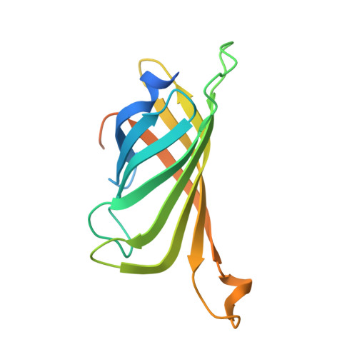
6ESS, 6ESU - PubMed Abstract:
Artificial metalloenzymes, resulting from incorporation of a metal cofactor within a host protein, have received increasing attention in the last decade. The directed evolution is presented of an artificial transfer hydrogenase (ATHase) based on the biotin-streptavidin technology using a straightforward procedure allowing screening in cell-free extracts. Two streptavidin isoforms were yielded with improved catalytic activity and selectivity for the reduction of cyclic imines. The evolved ATHases were stable under biphasic catalytic conditions. The X-ray structure analysis reveals that introducing bulky residues within the active site results in flexibility changes of the cofactor, thus increasing exposure of the metal to the protein surface and leading to a reversal of enantioselectivity. This hypothesis was confirmed by a multiscale approach based mostly on molecular dynamics and protein-ligand dockings.
- Department Chemistry, University of Basel, Mattenstrasse 24a, BPR 1096, Basel, 4002, Switzerland.
Organizational Affiliation: