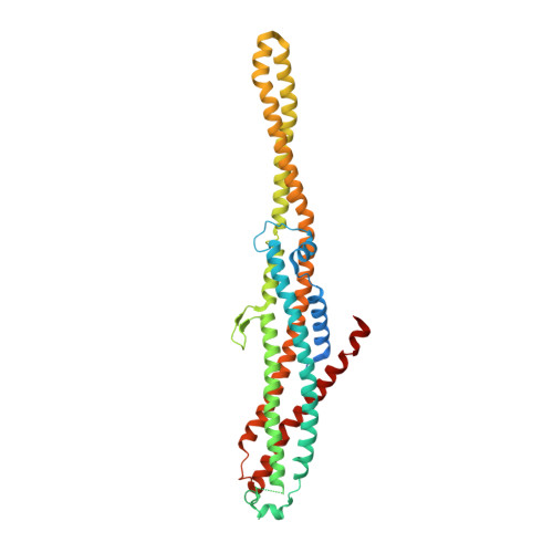
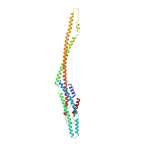
6EL1

YaxAB pore complex


Experimental Data Snapshot

  • Method: ELECTRON MICROSCOPY
  • Resolution: 6.10 Å
  • Aggregation State: PARTICLE 
  • Reconstruction Method: SINGLE PARTICLE 

Starting Models: experimental
View more details

wwPDB Validation   3D Report Full Report


This is version 1.2 of the entry. See complete history


Literature

Structure and mechanism of the two-component alpha-helical pore-forming toxin YaxAB.

Brauning, B.Bertosin, E.Praetorius, F.Ihling, C.Schatt, A.Adler, A.Richter, K.Sinz, A.Dietz, H.Groll, M.

(2018) Nat Commun 9: 1806-1806

  • DOI: https://doi.org/10.1038/s41467-018-04139-2
  • Primary Citation of Related Structures:  
    6EK4, 6EK7, 6EK8, 6EL1

  • PubMed Abstract: 

    Pore-forming toxins (PFT) are virulence factors that transform from soluble to membrane-bound states. The Yersinia YaxAB system represents a family of binary α-PFTs with orthologues in human, insect, and plant pathogens, with unknown structures. YaxAB was shown to be cytotoxic and likely involved in pathogenesis, though the molecular basis for its two-component lytic mechanism remains elusive. Here, we present crystal structures of YaxA and YaxB, together with a cryo-electron microscopy map of the YaxAB complex. Our structures reveal a pore predominantly composed of decamers of YaxA-YaxB heterodimers. Both subunits bear membrane-active moieties, but only YaxA is capable of binding to membranes by itself. YaxB can subsequently be recruited to membrane-associated YaxA and induced to present its lytic transmembrane helices. Pore formation can progress by further oligomerization of YaxA-YaxB dimers. Our results allow for a comparison between pore assemblies belonging to the wider ClyA-like family of α-PFTs, highlighting diverse pore architectures.


  • Organizational Affiliation

    Center for Integrated Protein Science Munich (CIPSM), Department of Chemistry, Chair of Biochemistry, Technische Universität München, Lichtenbergstrasse 4, 85747, Garching, Germany. bastian.braeuning@tum.de.


Macromolecules
Find similar proteins by:  (by identity cutoff)  |  3D Structure
Entity ID: 1
MoleculeChains Sequence LengthOrganismDetailsImage
YaxA410Yersinia enterocoliticaMutation(s): 0 
Gene Names: ERS137951_00706
Membrane Entity: Yes 
UniProt
Find proteins for A1JM51 (Yersinia enterocolitica serotype O:8 / biotype 1B (strain NCTC 13174 / 8081))
Explore A1JM51 
Go to UniProtKB:  A1JM51
Entity Groups  
Sequence Clusters30% Identity50% Identity70% Identity90% Identity95% Identity100% Identity
UniProt GroupA1JM51
Sequence Annotations
Expand
  • Reference Sequence
Find similar proteins by:  (by identity cutoff)  |  3D Structure
Entity ID: 2
MoleculeChains Sequence LengthOrganismDetailsImage
YaxB344Yersinia enterocoliticaMutation(s): 0 
Gene Names: YE1985
UniProt
Find proteins for A1JM52 (Yersinia enterocolitica serotype O:8 / biotype 1B (strain NCTC 13174 / 8081))
Explore A1JM52 
Go to UniProtKB:  A1JM52
Entity Groups  
Sequence Clusters30% Identity50% Identity70% Identity90% Identity95% Identity100% Identity
UniProt GroupA1JM52
Sequence Annotations
Expand
  • Reference Sequence
Experimental Data & Validation

Experimental Data

  • Method: ELECTRON MICROSCOPY
  • Resolution: 6.10 Å
  • Aggregation State: PARTICLE 
  • Reconstruction Method: SINGLE PARTICLE 
EM Software:
TaskSoftware PackageVersion
MODEL REFINEMENTPHENIX
RECONSTRUCTIONRELION

Structure Validation

View Full Validation Report


Entry History 

Deposition Data

Revision History  (Full details and data files)

  • Version 1.0: 2018-05-16
    Type: Initial release
  • Version 1.1: 2019-12-11
    Changes: Other
  • Version 1.2: 2024-05-15
    Changes: Data collection, Database references, Refinement description