6EJ7

Human Xylosyltransferase 1 in complex with UDP-xylose and peptide QEEEGAGGGQGG


Experimental Data Snapshot

  • Method: X-RAY DIFFRACTION
  • Resolution: 2.00 Å
  • R-Value Free: 0.230 
  • R-Value Work: 0.199 
  • R-Value Observed: 0.200 

wwPDB Validation   3D Report Full Report


Ligand Structure Quality Assessment 


This is version 1.3 of the entry. See complete history


Literature

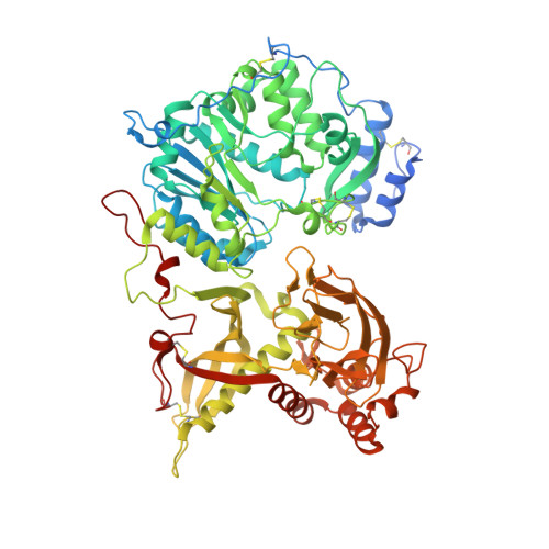
Structural Basis for the Initiation of Glycosaminoglycan Biosynthesis by Human Xylosyltransferase 1.

Briggs, D.C.Hohenester, E.

(2018) Structure 26: 801-809.e3

  • DOI: https://doi.org/10.1016/j.str.2018.03.014
  • Primary Citation of Related Structures:  
    6EJ7, 6EJ8, 6EJ9, 6EJA, 6EJB, 6EJC, 6EJD, 6EJE, 6FOA

  • PubMed Abstract: 

    Proteoglycans (PGs) are essential components of the animal extracellular matrix and are required for cell adhesion, migration, signaling, and immune function. PGs are composed of a core protein and long glycosaminoglycan (GAG) chains, which often specify PG function. GAG biosynthesis is initiated by peptide O-xylosyltransferases, which transfer xylose onto selected serine residues in the core proteins. We have determined crystal structures of human xylosyltransferase 1 (XT1) in complex with the sugar donor, UDP-xylose, and various acceptor peptides. The structures reveal unique active-site features that, in conjunction with functional experiments, explain the substrate specificity of XT1. A constriction within the peptide binding cleft requires the acceptor serine to be followed by glycine or alanine. The remainder of the cleft can accommodate a wide variety of sequences, but with a general preference for acidic residues. These findings provide a framework for understanding the selectivity of GAG attachment.


  • Organizational Affiliation

    Department of Life Sciences, Imperial College London, London SW7 2AZ, UK. Electronic address: david.briggs@crick.ac.uk.


Macromolecules
Find similar proteins by:  (by identity cutoff)  |  3D Structure
Entity ID: 1
MoleculeChains Sequence LengthOrganismDetailsImage
Xylosyltransferase 1751Homo sapiensMutation(s): 0 
Gene Names: XYLT1XT1
EC: 2.4.2.26
UniProt & NIH Common Fund Data Resources
Find proteins for Q86Y38 (Homo sapiens)
Explore Q86Y38 
Go to UniProtKB:  Q86Y38
PHAROS:  Q86Y38
GTEx:  ENSG00000103489 
Entity Groups  
Sequence Clusters30% Identity50% Identity70% Identity90% Identity95% Identity100% Identity
UniProt GroupQ86Y38
Sequence Annotations
Expand
  • Reference Sequence

Find similar proteins by:  Sequence   |   3D Structure  

Entity ID: 2
MoleculeChains Sequence LengthOrganismDetailsImage
Protein AMBP12Homo sapiensMutation(s): 3 
UniProt & NIH Common Fund Data Resources
Find proteins for P02760 (Homo sapiens)
Explore P02760 
Go to UniProtKB:  P02760
PHAROS:  P02760
GTEx:  ENSG00000106927 
Entity Groups  
Sequence Clusters30% Identity50% Identity70% Identity90% Identity95% Identity100% Identity
UniProt GroupP02760
Sequence Annotations
Expand
  • Reference Sequence
Small Molecules
Ligands 6 Unique
IDChains Name / Formula / InChI Key2D Diagram3D Interactions
UDX (Subject of Investigation/LOI)
Query on UDX

Download Ideal Coordinates CCD File 
D [auth A]URIDINE-5'-DIPHOSPHATE-XYLOPYRANOSE
C14 H22 N2 O16 P2
DQQDLYVHOTZLOR-OCIMBMBZSA-N
NAG
Query on NAG

Download Ideal Coordinates CCD File 
C [auth A]2-acetamido-2-deoxy-beta-D-glucopyranose
C8 H15 N O6
OVRNDRQMDRJTHS-FMDGEEDCSA-N
PEG
Query on PEG

Download Ideal Coordinates CCD File 
S [auth A],
T [auth A]
DI(HYDROXYETHYL)ETHER
C4 H10 O3
MTHSVFCYNBDYFN-UHFFFAOYSA-N
PO4
Query on PO4

Download Ideal Coordinates CCD File 
F [auth A]
G [auth A]
H [auth A]
I [auth A]
J [auth A]
F [auth A],
G [auth A],
H [auth A],
I [auth A],
J [auth A],
K [auth A],
L [auth A],
M [auth A],
N [auth A]
PHOSPHATE ION
O4 P
NBIIXXVUZAFLBC-UHFFFAOYSA-K
EDO
Query on EDO

Download Ideal Coordinates CCD File 
O [auth A],
P [auth A],
Q [auth A],
R [auth A]
1,2-ETHANEDIOL
C2 H6 O2
LYCAIKOWRPUZTN-UHFFFAOYSA-N
NA
Query on NA

Download Ideal Coordinates CCD File 
E [auth A]SODIUM ION
Na
FKNQFGJONOIPTF-UHFFFAOYSA-N
Experimental Data & Validation

Experimental Data

  • Method: X-RAY DIFFRACTION
  • Resolution: 2.00 Å
  • R-Value Free: 0.230 
  • R-Value Work: 0.199 
  • R-Value Observed: 0.200 
  • Space Group: P 21 21 21
Unit Cell:
Length ( Å )Angle ( ˚ )
a = 67.46α = 90
b = 86.89β = 90
c = 152.91γ = 90
Software Package:
Software NamePurpose
PHENIXrefinement
Aimlessdata scaling
PHASERphasing
PDB_EXTRACTdata extraction
xia2data reduction

Structure Validation

View Full Validation Report


Ligand Structure Quality Assessment 


Entry History & Funding Information

Deposition Data


Funding OrganizationLocationGrant Number
Wellcome TrustUnited Kingdom101748/Z/13/Z

Revision History  (Full details and data files)

  • Version 1.0: 2018-05-02
    Type: Initial release
  • Version 1.1: 2018-06-13
    Changes: Data collection, Database references
  • Version 1.2: 2020-07-29
    Type: Remediation
    Reason: Carbohydrate remediation
    Changes: Data collection, Derived calculations, Structure summary
  • Version 1.3: 2024-01-17
    Changes: Data collection, Database references, Refinement description, Structure summary