Engineering methionine gamma-lyase from Citrobacter freundii for anticancer activity.
Raboni, S., Revtovich, S., Demitri, N., Giabbai, B., Storici, P., Cocconcelli, C., Faggiano, S., Rosini, E., Pollegioni, L., Galati, S., Buschini, A., Morozova, E., Kulikova, V., Nikulin, A., Gabellieri, E., Cioni, P., Demidkina, T., Mozzarelli, A.(2018) Biochim Biophys Acta Proteins Proteom 1866: 1260-1270
- PubMed: 30268810 Search on PubMed
- DOI: https://doi.org/10.1016/j.bbapap.2018.09.011
- Primary Citation Related Structures:
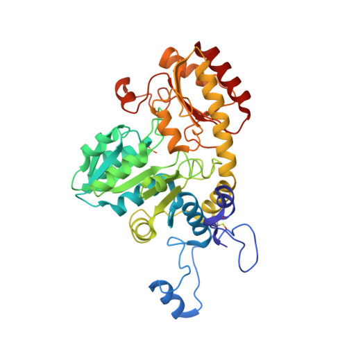
6EGR - PubMed Abstract:
Methionine deprivation of cancer cells, which are deficient in methionine biosynthesis, has been envisioned as a therapeutic strategy to reduce cancer cell viability. Methionine γ-lyase (MGL), an enzyme that degrades methionine, has been exploited to selectively remove the amino acid from cancer cell environment. In order to increase MGL catalytic activity, we performed sequence and structure conservation analysis of MGLs from various microorganisms. Whereas most of the residues in the active site and at the dimer interface were found to be conserved, residues located in the C-terminal flexible loop, forming a wall of the active site entry channel, were found to be variable. Therefore, we carried out site-saturation mutagenesis at four independent positions of the C-terminal flexible loop, P357, V358, P360 and A366 of MGL from Citrobacter freundii, generating libraries that were screened for activity. Among the active variants, V358Y exhibits a 1.9-fold increase in the catalytic rate and a 3-fold increase in K M , resulting in a catalytic efficiency similar to wild type MGL. V358Y cytotoxic activity was assessed towards a panel of cancer and nonmalignant cell lines and found to exhibit IC 50 lower than the wild type. The comparison of the 3D-structure of V358Y MGL with other MGL available structures indicates that the C-terminal loop is either in an open or closed conformation that does not depend on the amino acid at position 358. Nevertheless, mutations at this position allosterically affects catalysis.
- Department of Food and Drug, University of Parma, Parma, Italy; Institute of Biophysics, National Research Council, Pisa, Italy.
Organizational Affiliation: