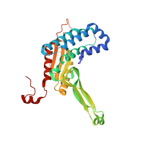
6EG7

BbvCI B2 dimer with I3C clusters


Experimental Data Snapshot

  • Method: X-RAY DIFFRACTION
  • Resolution: 3.00 Å
  • R-Value Free: 0.262 
  • R-Value Work: 0.213 
  • R-Value Observed: 0.216 

wwPDB Validation   3D Report Full Report


Ligand Structure Quality Assessment 


This is version 1.4 of the entry. See complete history


Literature

Structure, subunit organization and behavior of the asymmetric Type IIT restriction endonuclease BbvCI.

Shen, B.W.Doyle, L.Bradley, P.Heiter, D.F.Lunnen, K.D.Wilson, G.G.Stoddard, B.L.

(2019) Nucleic Acids Res 47: 450-467

  • DOI: https://doi.org/10.1093/nar/gky1059
  • Primary Citation of Related Structures:  
    6EG7, 6M9G, 6MAF, 6MAG

  • PubMed Abstract: 

    BbvCI, a Type IIT restriction endonuclease, recognizes and cleaves the seven base pair sequence 5'-CCTCAGC-3', generating 3-base, 5'-overhangs. BbvCI is composed of two protein subunits, each containing one catalytic site. Either site can be inactivated by mutation resulting in enzyme variants that nick DNA in a strand-specific manner. Here we demonstrate that the holoenzyme is labile, with the R1 subunit dissociating at low pH. Crystallization of the R2 subunit under such conditions revealed an elongated dimer with the two catalytic sites located on opposite sides. Subsequent crystallization at physiological pH revealed a tetramer comprising two copies of each subunit, with a pair of deep clefts each containing two catalytic sites appropriately positioned and oriented for DNA cleavage. This domain organization was further validated with single-chain protein constructs in which the two enzyme subunits were tethered via peptide linkers of variable length. We were unable to crystallize a DNA-bound complex; however, structural similarity to previously crystallized restriction endonucleases facilitated creation of an energy-minimized model bound to DNA, and identification of candidate residues responsible for target recognition. Mutation of residues predicted to recognize the central C:G base pair resulted in an altered enzyme that recognizes and cleaves CCTNAGC (N = any base).


  • Organizational Affiliation

    Division of Basic Sciences, Fred Hutchinson Cancer Research Center, 1100 Fairview Ave. N., Seattle, WA 98109, USA.


Macromolecules
Find similar proteins by:  (by identity cutoff)  |  3D Structure
Entity ID: 1
MoleculeChains Sequence LengthOrganismDetailsImage
BbvCI endonuclease subunit 2
A, B
285Brevibacillus brevisMutation(s): 0 
Gene Names: bbvCIR-2
UniProt
Find proteins for Q5D6Y4 (Brevibacillus brevis)
Explore Q5D6Y4 
Go to UniProtKB:  Q5D6Y4
Entity Groups  
Sequence Clusters30% Identity50% Identity70% Identity90% Identity95% Identity100% Identity
UniProt GroupQ5D6Y4
Sequence Annotations
Expand
  • Reference Sequence
Small Molecules
Ligands 3 Unique
IDChains Name / Formula / InChI Key2D Diagram3D Interactions
I3C
Query on I3C

Download Ideal Coordinates CCD File 
G [auth A]
H [auth A]
I [auth A]
J [auth A]
L [auth B]
G [auth A],
H [auth A],
I [auth A],
J [auth A],
L [auth B],
M [auth B],
N [auth B],
O [auth B]
5-amino-2,4,6-triiodobenzene-1,3-dicarboxylic acid
C8 H4 I3 N O4
JEZJSNULLBSYHV-UHFFFAOYSA-N
PEG
Query on PEG

Download Ideal Coordinates CCD File 
E [auth A],
K [auth B]
DI(HYDROXYETHYL)ETHER
C4 H10 O3
MTHSVFCYNBDYFN-UHFFFAOYSA-N
EDO
Query on EDO

Download Ideal Coordinates CCD File 
C [auth A],
D [auth A],
F [auth A]
1,2-ETHANEDIOL
C2 H6 O2
LYCAIKOWRPUZTN-UHFFFAOYSA-N
Experimental Data & Validation

Experimental Data

  • Method: X-RAY DIFFRACTION
  • Resolution: 3.00 Å
  • R-Value Free: 0.262 
  • R-Value Work: 0.213 
  • R-Value Observed: 0.216 
  • Space Group: C 2 2 21
Unit Cell:
Length ( Å )Angle ( ˚ )
a = 93.888α = 90
b = 168.245β = 90
c = 106.477γ = 90
Software Package:
Software NamePurpose
REFMACrefinement
HKL-2000data reduction
HKL-2000data scaling
SHELXDEphasing

Structure Validation

View Full Validation Report


Ligand Structure Quality Assessment 


Entry History & Funding Information

Deposition Data


Funding OrganizationLocationGrant Number
National Institutes of Health/National Human Genome Research Institute (NIH/NHGRI)United StatesR01 GM105691 to BLS

Revision History  (Full details and data files)

  • Version 1.0: 2018-11-14
    Type: Initial release
  • Version 1.1: 2018-11-21
    Changes: Data collection, Database references
  • Version 1.2: 2019-01-23
    Changes: Data collection, Database references
  • Version 1.3: 2019-12-18
    Changes: Author supporting evidence
  • Version 1.4: 2024-03-13
    Changes: Data collection, Database references, Refinement description