


Structure-based identification of uncompetitive inhibitors for Staphylococcus aureus pantothenate kinase.
Chen, Y., Antoshchenko, T., Strauss, E., Barnard, L., Huang, Y.H.To be published.
Experimental Data Snapshot
Entity ID: 1 | |||||
|---|---|---|---|---|---|
| Molecule | Chains | Sequence Length | Organism | Details | Image |
| Type II pantothenate kinase | 267 | Staphylococcus aureus | Mutation(s): 0 Gene Names: coaW, BTN44_13005, C3B39_13135, EP54_10775, EQ90_06670, HMPREF3211_02599 EC: 2.7.1.33 | 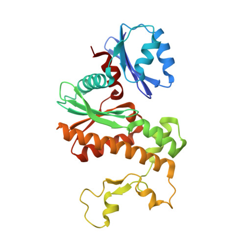 | |
UniProt | |||||
Entity Groups | |||||
| Sequence Clusters | 30% Identity50% Identity70% Identity90% Identity95% Identity100% Identity | ||||
| UniProt Group | Q2FWC7 | ||||
Sequence AnnotationsExpand | |||||
Reference Sequence | |||||
| Ligands 2 Unique | |||||
|---|---|---|---|---|---|
| ID | Chains | Name / Formula / InChI Key | 2D Diagram | 3D Interactions | |
| ADP Download:Ideal Coordinates CCD File | E [auth A], G [auth B], I [auth C], K [auth D] | ADENOSINE-5'-DIPHOSPHATE C10 H15 N5 O10 P2 XTWYTFMLZFPYCI-KQYNXXCUSA-N |  | ||
| 27Q Download:Ideal Coordinates CCD File | F [auth A], H [auth B], J [auth C], L [auth D] | N-heptyl-N~3~-[(2R)-2-hydroxy-3,3-dimethyl-4-(phosphonooxy)butanoyl]-beta-alaninamide C16 H33 N2 O7 P QTSWMJJVYAHWSJ-AWEZNQCLSA-N |  | ||
| Length ( Å ) | Angle ( ˚ ) |
|---|---|
| a = 72.8 | α = 90 |
| b = 56.72 | β = 99.81 |
| c = 133.62 | γ = 90 |
| Software Name | Purpose |
|---|---|
| REFMAC | refinement |
| PDB_EXTRACT | data extraction |
| XDS | data reduction |
| SCALA | data scaling |
| BUSTER | phasing |


