


Crystal structure of tryptophan synthase from M. tuberculosis - closed form with BRD6309 bound
Chang, C., Michalska, K., Maltseva, N.I., Jedrzejczak, R., McCarren, P., Nag, P.P., Joachimiak, A., Satchell, K.To be published.
Experimental Data Snapshot
Starting Model: experimental
View more details
Entity ID: 1 | |||||
|---|---|---|---|---|---|
| Molecule | Chains | Sequence Length | Organism | Details | Image |
| Tryptophan synthase alpha chain | A, C [auth G], E, G [auth C] | 276 | Mycobacterium tuberculosis H37Rv | Mutation(s): 0 Gene Names: trpA, Rv1613, MTCY01B2.05 EC: 4.2.1.20 | 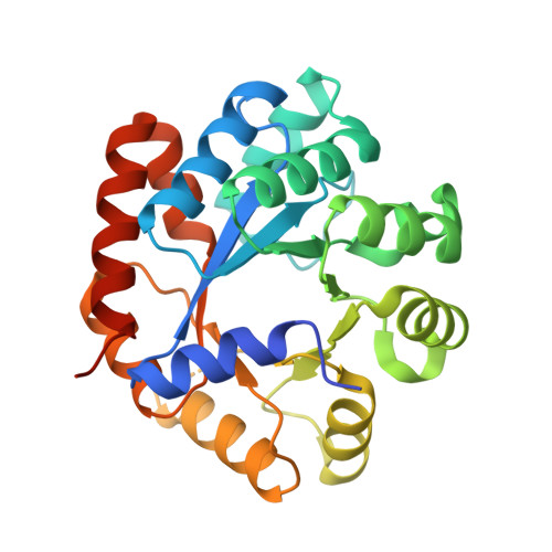 |
UniProt | |||||
Entity Groups | |||||
| Sequence Clusters | 30% Identity50% Identity70% Identity90% Identity95% Identity100% Identity | ||||
| UniProt Group | P9WFY1 | ||||
Sequence AnnotationsExpand | |||||
Reference Sequence | |||||
Entity ID: 2 | |||||
|---|---|---|---|---|---|
| Molecule | Chains | Sequence Length | Organism | Details | Image |
| Tryptophan synthase beta chain | B, D [auth H], F, H [auth D] | 410 | Mycobacterium tuberculosis H37Rv | Mutation(s): 0 Gene Names: trpB, Rv1612, MTCY01B2.04 EC: 4.2.1.20 | 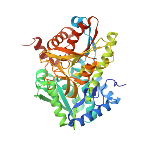 |
UniProt | |||||
Entity Groups | |||||
| Sequence Clusters | 30% Identity50% Identity70% Identity90% Identity95% Identity100% Identity | ||||
| UniProt Group | P9WFX9 | ||||
Sequence AnnotationsExpand | |||||
Reference Sequence | |||||
| Ligands 9 Unique | |||||
|---|---|---|---|---|---|
| ID | Chains | Name / Formula / InChI Key | 2D Diagram | 3D Interactions | |
| P1T Download:Ideal Coordinates CCD File | CA [auth H], FB [auth D], L [auth B], OA [auth F] | 2-[({3-HYDROXY-2-METHYL-5-[(PHOSPHONOOXY)METHYL]PYRIDIN-4-YL}METHYL)AMINO]ACRYLIC ACID C11 H15 N2 O7 P BXUDKFHCAMQSRX-UHFFFAOYSA-N |  | ||
| HDJ Download:Ideal Coordinates CCD File | AB [auth F], JA [auth H], PB [auth D], V [auth B] | (2R,3S,4R)-3-(2',6'-difluoro-4'-methyl[1,1'-biphenyl]-4-yl)-4-(fluoromethyl)azetidine-2-carbonitrile C18 H15 F3 N2 OZFKPHFHTNQIQS-XYJFISCASA-N |  | ||
| PGE Download:Ideal Coordinates CCD File | QB [auth D] | TRIETHYLENE GLYCOL C6 H14 O4 ZIBGPFATKBEMQZ-UHFFFAOYSA-N |  | ||
| CS Download:Ideal Coordinates CCD File | DA [auth H] GB [auth D] HB [auth D] M [auth B] N [auth B] | CESIUM ION Cs NCMHKCKGHRPLCM-UHFFFAOYSA-N |  | ||
| MLA Download:Ideal Coordinates CCD File | CB [auth F], MA [auth H] | MALONIC ACID C3 H4 O4 OFOBLEOULBTSOW-UHFFFAOYSA-N |  | ||
| MLI Download:Ideal Coordinates CCD File | DB [auth C], I [auth A], NA [auth E], X [auth G] | MALONATE ION C3 H2 O4 OFOBLEOULBTSOW-UHFFFAOYSA-L |  | ||
| EDO Download:Ideal Coordinates CCD File | BB [auth F] | 1,2-ETHANEDIOL C2 H6 O2 LYCAIKOWRPUZTN-UHFFFAOYSA-N |  | ||
| ACT Download:Ideal Coordinates CCD File | KA [auth H], LA [auth H], W [auth B] | ACETATE ION C2 H3 O2 QTBSBXVTEAMEQO-UHFFFAOYSA-M |  | ||
| FMT Download:Ideal Coordinates CCD File | AA [auth G] BA [auth G] EA [auth H] EB [auth C] FA [auth H] | FORMIC ACID C H2 O2 BDAGIHXWWSANSR-UHFFFAOYSA-N |  | ||
| Length ( Å ) | Angle ( ˚ ) |
|---|---|
| a = 135.093 | α = 90 |
| b = 159.396 | β = 90 |
| c = 165.226 | γ = 90 |
| Software Name | Purpose |
|---|---|
| SCALEPACK | data scaling |
| PHENIX | refinement |
| PDB_EXTRACT | data extraction |
| HKL-3000 | data reduction |
| HKL-3000 | phasing |


