6DHW | pdb_00006dhw

Crystal structure of primase iron-sulfur domain (266-457)


Experimental Data Snapshot

  • Method: X-RAY DIFFRACTION
  • Resolution: 2.01 Å
  • R-Value Free: 
    0.211 (Depositor), 0.210 (DCC) 
  • R-Value Work: 
    0.181 (Depositor), 0.181 (DCC) 
  • R-Value Observed: 
    0.182 (Depositor) 

Starting Model: experimental
View more details

wwPDB Validation 3D Report Full Report

Validation slider image for 6DHW

This is version 1.4 of the entry. See complete history

Literature

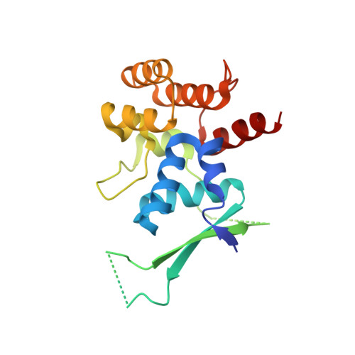
Functional and structural similarity of human DNA primase [4Fe4S] cluster domain constructs.

Holt, M.E.Salay, L.E.O'Brien, E.Barton, J.K.Chazin, W.J.

(2018) PLoS One 13: e0209345-e0209345

  • DOI: https://doi.org/10.1371/journal.pone.0209345
  • Primary Citation Related Structures: 
    6DHW

  • PubMed Abstract: 

    The regulatory subunit of human DNA primase has a C-terminal domain (p58C) that contains a [4Fe4S] cluster and binds DNA. Previous electrochemical analysis of a p58C construct revealed that its affinity for DNA is sensitive to the redox state of the [4Fe4S] cluster. Concerns about the validity of this conclusion have been raised, based in part on differences in X-ray crystal structures of the p58C272-464 construct used for that study and that of a N-terminally shifted p58C266-456 construct and consequently, an assumption that p58C272-464 has abnormal physical and functional properties. To address this controversy, a new p58C266-464 construct containing all residues was crystallized under the conditions previously used for crystallizing p58C272-464, and the solution structures of both constructs were assessed using circular dichroism and NMR spectroscopy. In the new crystal structure, p58C266-464 exhibits the same elements of secondary structure near the DNA binding site as observed in the crystal structure of p58C272-464. Moreover, in solution, circular dichroism and 15N,1H-heteronuclear single quantum coherence (HSQC) NMR spectra show there are no significant differences in the distribution of secondary structures or in the tertiary structure or the two constructs. To validate that the two constructs have the same functional properties, binding of a primed DNA template was measured using a fluorescence-based DNA binding assay, and the affinities for this substrate were the same (3.4 ± 0.5 μM and 2.7 ± 0.3 μM, respectively). The electrochemical properties of p58C266-464 were also measured and this p58C construct was able to engage in redox switching on DNA with the same efficiency as p58C272-464. Together, these results show that although p58C can be stabilized in different conformations in the crystalline state, in solution there is effectively no difference in the structure and functional properties of p58C constructs of different lengths.


  • Organizational Affiliation
    • Departments of Biochemistry and Chemistry, and Center for Structural Biology, Vanderbilt University, Nashville, Tennessee, United States of America.

Macromolecule Content 

  • Total Structure Weight: 45.45 kDa 
  • Atom Count: 2,948 
  • Modeled Residue Count: 336 
  • Deposited Residue Count: 384 
  • Unique protein chains: 1

Macromolecules

Find similar proteins by:|  3D Structure
Entity ID: 1
MoleculeChains  Sequence LengthOrganismDetailsImage
DNA primase large subunit
A, B
192Homo sapiensMutation(s): 0 
Gene Names: PRIM2PRIM2A
EC: 2.7.7
UniProt & NIH Common Fund Data Resources
Find proteins for P49643 (Homo sapiens)
Explore P49643 
Go to UniProtKB:  P49643
PHAROS:  P49643
GTEx:  ENSG00000146143 
Entity Groups
Sequence Clusters30% Identity50% Identity70% Identity90% Identity95% Identity100% Identity
UniProt GroupP49643
Sequence Annotations
Expand
Reference Sequence

Small Molecules

Ligands 2 Unique
IDChains Name / Formula / InChI Key2D Diagram3D Interactions
SF4

Query on SF4


Download:Ideal Coordinates CCD File
C [auth A],
F [auth B]
IRON/SULFUR CLUSTER
Fe4 S4
LJBDFODJNLIPKO-UHFFFAOYSA-N
SO4

Query on SO4


Download:Ideal Coordinates CCD File
D [auth A]
E [auth A]
G [auth B]
H [auth B]
I [auth B]
D [auth A],
E [auth A],
G [auth B],
H [auth B],
I [auth B],
J [auth B]
SULFATE ION
O4 S
QAOWNCQODCNURD-UHFFFAOYSA-L

Experimental Data & Validation

Experimental Data

  • Method: X-RAY DIFFRACTION
  • Resolution: 2.01 Å
  • R-Value Free:  0.211 (Depositor), 0.210 (DCC) 
  • R-Value Work:  0.181 (Depositor), 0.181 (DCC) 
  • R-Value Observed: 0.182 (Depositor) 
Space Group: C 1 2 1
Unit Cell:
Length ( Å )Angle ( ˚ )
a = 110.193α = 90
b = 52.56β = 115.08
c = 88.774γ = 90
Software Package:
Software NamePurpose
PHENIXrefinement
DENZOdata reduction
HKL-2000data scaling
PHASERphasing
PDB_EXTRACTdata extraction

Structure Validation

View Full Validation Report


Entry History 

& Funding Information

Deposition Data


Funding OrganizationLocationGrant Number
National Institutes of Health/National Institute of General Medical Sciences (NIH/NIGMS)United StatesR35 GM118089

Revision History  (Full details and data files)

  • Version 1.0: 2018-12-19
    Type: Initial release
  • Version 1.1: 2019-01-02
    Changes: Data collection, Database references
  • Version 1.2: 2020-01-01
    Changes: Author supporting evidence
  • Version 1.3: 2023-10-11
    Changes: Data collection, Database references, Refinement description
  • Version 1.4: 2024-11-06
    Changes: Structure summary