6DHC | pdb_00006dhc

X-ray structure of BACE1 in complex with a bicyclic isoxazoline carboxamide as the P3 ligand

  • Classification: HYDROLASE
  • Organism(s): Homo sapiens
  • Expression System: Escherichia coli
  • Mutation(s): No 

  • Deposited: 2018-05-19 Released: 2018-07-25 
  • Deposition Author(s): Mesecar, A.D., Lendy, E.K.
  • Funding Organization(s): National Institutes of Health/National Institute of General Medical Sciences (NIH/NIGMS)

Experimental Data Snapshot

  • Method: X-RAY DIFFRACTION
  • Resolution: 2.85 Å
  • R-Value Free: 
    0.218 (Depositor), 0.221 (DCC) 
  • R-Value Work: 
    0.172 (Depositor), 0.175 (DCC) 
  • R-Value Observed: 
    0.174 (Depositor) 

wwPDB Validation 3D Report Full Report

Validation slider image for 6DHC

Ligand Structure Quality Assessment 


This is version 1.3 of the entry. See complete history

Literature

Design, synthesis, X-ray studies, and biological evaluation of novel BACE1 inhibitors with bicyclic isoxazoline carboxamides as the P3 ligand.

Ghosh, A.K.Ghosh, K.Brindisi, M.Lendy, E.K.Yen, Y.C.Kumaragurubaran, N.Huang, X.Tang, J.Mesecar, A.D.

(2018) Bioorg Med Chem Lett 28: 2605-2610

  • DOI: https://doi.org/10.1016/j.bmcl.2018.06.045
  • Primary Citation Related Structures: 
    6DHC

  • PubMed Abstract: 

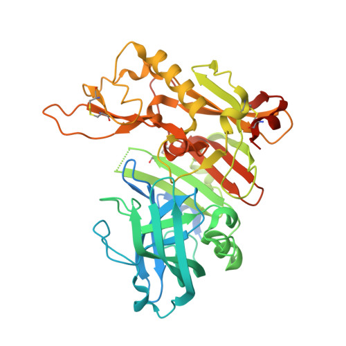
    We describe the design, synthesis, X-ray studies, and biological evaluation of novel BACE1 inhibitors containing bicyclic isoxazoline carboxamides as the P3 ligand in combination with methyl cysteine, methylsulfonylalanine and Boc-amino alanine as P2 ligands. Inhibitor 3a displayed a BACE1 K i value of 10.9 nM and EC 50 of 343 nM. The X-ray structure of 3a bound to the active site of BACE1 was determined at 2.85 Å resolution. The structure revealed that the major molecular interactions between BACE1 and the bicyclic tetrahydrofuranyl isoxazoline heterocycle are van der Waals in nature.


  • Organizational Affiliation
    • Department of Chemistry, Purdue University, West Lafayette, IN 47907, United States; Department of Medicinal Chemistry, Purdue University, West Lafayette, IN 47907, United States. Electronic address: akghosh@purdue.edu.

Macromolecule Content 

  • Total Structure Weight: 149.53 kDa 
  • Atom Count: 9,364 
  • Modeled Residue Count: 1,135 
  • Deposited Residue Count: 1,323 
  • Unique protein chains: 1

Macromolecules

Find similar proteins by:|  3D Structure
Entity ID: 1
MoleculeChains  Sequence LengthOrganismDetailsImage
Beta-secretase 1
A, B, C
441Homo sapiensMutation(s): 0 
Gene Names: BACE1BACEKIAA1149
EC: 3.4.23.46
UniProt & NIH Common Fund Data Resources
Find proteins for P56817 (Homo sapiens)
Explore P56817 
Go to UniProtKB:  P56817
PHAROS:  P56817
GTEx:  ENSG00000186318 
Entity Groups
Sequence Clusters30% Identity50% Identity70% Identity90% Identity95% Identity100% Identity
UniProt GroupP56817
Sequence Annotations
Expand
Reference Sequence

Small Molecules

Ligands 4 Unique
IDChains Name / Formula / InChI Key2D Diagram3D Interactions
GHJ
(Subject of Investigation/LOI)

Query on GHJ


Download:Ideal Coordinates CCD File
D [auth A],
K [auth B],
P [auth C]
(3R,3aR,6aS)-N-[(4R,7S,8S,10R,13S)-8-hydroxy-10,17-dimethyl-7-(2-methylpropyl)-5,11,14-trioxo-13-(propan-2-yl)-2-thia-6,12,15-triazaoctadecan-4-yl]hexahydrofuro[3,2-d][1,2]oxazole-3-carboxamide
C29 H53 N5 O7 S
FHZUZMVZKPPWHG-VRUQXOILSA-N
SO4

Query on SO4


Download:Ideal Coordinates CCD File
T [auth C]SULFATE ION
O4 S
QAOWNCQODCNURD-UHFFFAOYSA-L
GOL

Query on GOL


Download:Ideal Coordinates CCD File
E [auth A]
F [auth A]
G [auth A]
L [auth B]
M [auth B]
E [auth A],
F [auth A],
G [auth A],
L [auth B],
M [auth B],
N [auth B],
Q [auth C],
R [auth C],
S [auth C]
GLYCEROL
C3 H8 O3
PEDCQBHIVMGVHV-UHFFFAOYSA-N
URE

Query on URE


Download:Ideal Coordinates CCD File
H [auth A],
I [auth A],
J [auth A],
O [auth B]
UREA
C H4 N2 O
XSQUKJJJFZCRTK-UHFFFAOYSA-N

Experimental Data & Validation

Experimental Data

  • Method: X-RAY DIFFRACTION
  • Resolution: 2.85 Å
  • R-Value Free:  0.218 (Depositor), 0.221 (DCC) 
  • R-Value Work:  0.172 (Depositor), 0.175 (DCC) 
  • R-Value Observed: 0.174 (Depositor) 
Space Group: P 1 21 1
Unit Cell:
Length ( Å )Angle ( ˚ )
a = 81.987α = 90
b = 103.322β = 102.98
c = 100.959γ = 90
Software Package:
Software NamePurpose
PHENIXrefinement
MOSFLMdata reduction
SCALAdata scaling
PHENIXphasing

Structure Validation

View Full Validation Report


Ligand Structure Quality Assessment 


Entry History 

& Funding Information

Deposition Data


Funding OrganizationLocationGrant Number
National Institutes of Health/National Institute of General Medical Sciences (NIH/NIGMS)United StatesGM053386

Revision History  (Full details and data files)

  • Version 1.0: 2018-07-25
    Type: Initial release
  • Version 1.1: 2018-08-22
    Changes: Data collection, Database references
  • Version 1.2: 2020-01-01
    Changes: Author supporting evidence
  • Version 1.3: 2024-10-23
    Changes: Data collection, Database references, Structure summary