Crystal-on-crystal chips for in situ serial diffraction at room temperature.
Ren, Z., Ayhan, M., Bandara, S., Bowatte, K., Kumarapperuma, I., Gunawardana, S., Shin, H., Wang, C., Zeng, X., Yang, X.(2018) Lab Chip 18: 2246-2256
- PubMed: 29952383 Search on PubMedSearch on PubMed Central
- DOI: https://doi.org/10.1039/c8lc00489g
- Primary Citation Related Structures:
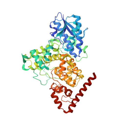
6DD6, 6DD7 - PubMed Abstract:
Recent developments in serial crystallography at X-ray free electron lasers (XFELs) and synchrotrons have been driven by two scientific goals in structural biology - first, static structure determination from nano or microcrystals of membrane proteins and large complexes that are difficult for conventional cryocrystallography, and second, direct observations of transient structural species in biochemical reactions at near atomic resolution. Since room-temperature diffraction experiments naturally demand a large quantity of purified protein, sample economy is critically important for all steps of serial crystallography from crystallization, crystal delivery to data collection. Here we report the development and applications of "crystal-on-crystal" devices to facilitate large-scale in situ serial diffraction experiments on protein crystals of all sizes - large, small, or microscopic. We show that the monocrystalline quartz as a substrate material prevents vapor loss during crystallization and significantly reduces background X-ray scattering. These devices can be readily adopted at XFEL and synchrotron beamlines, which enable efficient delivery of hundreds to millions of crystals to the X-ray beam, with an overall protein consumption per dataset comparable to that of cryocrystallography.
- Department of Chemistry, The University of Illinois at Chicago, Chicago, IL 60607, USA. zren@uic.edu xiaojing@uic.edu.
Organizational Affiliation: