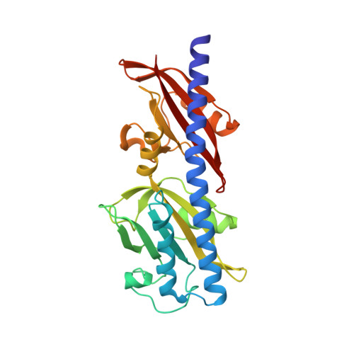
Structure of the sensory domain of McpX fromSinorhizobium meliloti, the first known bacterial chemotactic sensor for quaternary ammonium compounds.
Shrestha, M., Compton, K.K., Mancl, J.M., Webb, B.A., Brown, A.M., Scharf, B.E., Schubot, F.D.(2018) Biochem J 475: 3949-3962
- PubMed: 30442721 Search on PubMed
- DOI: https://doi.org/10.1042/BCJ20180769
- Primary Citation Related Structures:
6D8V - PubMed Abstract:
The α-proteobacterium Sinorhizobium meliloti can live freely in the soil or engage in a symbiosis with its legume host. S. meliloti facilitates nitrogen fixation in root nodules, thus providing pivotal, utilizable nitrogen to the host. The organism has eight chemoreceptors, namely McpT to McpZ and IcpA that facilitate chemotaxis. McpX is the first known bacterial sensor of quaternary ammonium compounds (QACs) such as choline and betaines. Because QACs are exuded at chemotaxis-relevant concentrations by germinating alfalfa seeds, McpX has been proposed to contribute to host-specific chemotaxis. We have determined the crystal structure of the McpX periplasmic region (McpX PR ) in complex with the proline betaine at 2.7 Å resolution. In the crystal, the protein forms a symmetric dimer with one proline betaine molecule bound to each monomer of McpX PR within membrane-distal CACHE module. The ligand is bound through cation-πinteractions with four aromatic amino acid residues. Mutational analysis in conjunction with binding studies revealed that a conserved aspartate residue is pivotal for ligand binding. We discovered that, in a striking example of convergent evolution, the ligand-binding site of McpX PR resembles that of a group of structurally unrelated betaine-binding proteins including ProX and OpuAC. Through this comparison and docking studies, we rationalized the specificity of McpX PR for this specific group of ligands. Collectively, our structural, biochemical, and molecular docking data have revealed the molecular determinants in McpX that are crucial for its rare ligand specificity for QACs.
- Department of Biological Sciences, Virginia Tech, Derring Hall, Blacksburg, VA 24061, U.S.A.
Organizational Affiliation: