6D54

Low Temperature joint X-ray/neutron structure of DNA oligonucleotide d(GTGGCCAC)2 with 2'-SeCH3 modification on Cyt5


Experimental Data Snapshot

  • Method: X-RAY DIFFRACTION
  • Resolution: 1.65 Å
  • R-Value Free: 0.246 
  • R-Value Work: 0.230 

  • Method: NEUTRON DIFFRACTION
  • Resolution: 1.90 Å
  • R-Value Free: 0.260 
  • R-Value Work: 0.234 

wwPDB Validation   3D Report Full Report


This is version 2.1 of the entry. See complete history


Literature

Temperature-Induced Replacement of Phosphate Proton with Metal Ion Captured in Neutron Structures of A-DNA.

Vandavasi, V.G.Blakeley, M.P.Keen, D.A.Hu, L.R.Huang, Z.Kovalevsky, A.

(2018) Structure 26: 1645

  • DOI: https://doi.org/10.1016/j.str.2018.08.001
  • Primary Citation of Related Structures:  
    6D4L, 6D54

  • PubMed Abstract: 

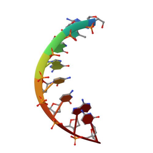
    Nucleic acids can fold into well-defined 3D structures that help determine their function. Knowing precise nucleic acid structures can also be used for the design of nucleic acid-based therapeutics. However, locations of hydrogen atoms, which are key players of nucleic acid function, are normally not determined with X-ray crystallography. Accurate determination of hydrogen atom positions can provide indispensable information on protonation states, hydrogen bonding, and water architecture in nucleic acids. Here, we used neutron crystallography in combination with X-ray diffraction to obtain joint X-ray/neutron structures at both room and cryo temperatures of a self-complementary A-DNA oligonucleotide d[GTGG(C Se )CAC] 2 containing 2'-SeCH 3 modification on Cyt5 (C Se ) at pH 5.6. We directly observed protonation of a backbone phosphate oxygen of Ade7 at room temperature. The proton is replaced with hydrated Mg 2+ upon cooling the crystal to 100 K, indicating that metal binding is favored at low temperature, whereas proton binding is dominant at room temperature.


  • Organizational Affiliation

    Neutron Scattering Division, Neutron Sciences Directorate, Oak Ridge National Laboratory, Oak Ridge, TN 37922, USA.


Macromolecules

Find similar nucleic acids by:  Sequence   |   3D Structure  

Entity ID: 1
MoleculeChains LengthOrganismImage
DNA (5'-D(*GP*TP*GP*GP*(CSL)P*CP*AP*C)-3')8synthetic construct
Sequence Annotations
Expand
  • Reference Sequence
Small Molecules
Ligands 1 Unique
IDChains Name / Formula / InChI Key2D Diagram3D Interactions
MG
Query on MG

Download Ideal Coordinates CCD File 
B [auth A],
C [auth A]
MAGNESIUM ION
Mg
JLVVSXFLKOJNIY-UHFFFAOYSA-N
Experimental Data & Validation

Experimental Data

  • Method: X-RAY DIFFRACTION
  • Resolution: 1.65 Å
  • R-Value Free: 0.246 
  • R-Value Work: 0.230 
  • Space Group: P 43 21 2
  • Method: NEUTRON DIFFRACTION
  • Resolution: 1.90 Å
  • R-Value Free: 0.260 
  • R-Value Work: 0.234 
  • Space Group: P 43 21 2
Unit Cell:
Length ( Å )Angle ( ˚ )
a = 41.884α = 90
b = 41.884β = 90
c = 24.307γ = 90
Software Package:
Software NamePurpose
nCNSrefinement
HKL-3000data reduction
HKL-3000data scaling
PHASERphasing
LAUEGENdata reduction
LSCALEdata scaling
CNSphasing

Structure Validation

View Full Validation Report


Entry History 

Deposition Data

Revision History  (Full details and data files)

  • Version 1.0: 2018-10-17
    Type: Initial release
  • Version 1.1: 2018-12-19
    Changes: Data collection, Database references
  • Version 2.0: 2022-03-02
    Changes: Advisory, Data collection, Database references, Derived calculations, Polymer sequence, Refinement description, Structure summary
  • Version 2.1: 2023-10-04
    Changes: Data collection, Refinement description