6D32

Crystal structure of Xenopus Smoothened in complex with cyclopamine


Experimental Data Snapshot

  • Method: X-RAY DIFFRACTION
  • Resolution: 3.75 Å
  • R-Value Free: 0.260 
  • R-Value Work: 0.227 
  • R-Value Observed: 0.229 

wwPDB Validation   3D Report Full Report


Ligand Structure Quality Assessment 


This is version 1.4 of the entry. See complete history


Literature

Structural Basis of Smoothened Activation in Hedgehog Signaling.

Huang, P.Zheng, S.Wierbowski, B.M.Kim, Y.Nedelcu, D.Aravena, L.Liu, J.Kruse, A.C.Salic, A.

(2018) Cell 174: 312-324.e16

  • DOI: https://doi.org/10.1016/j.cell.2018.04.029
  • Primary Citation of Related Structures:  
    6D32, 6D35

  • PubMed Abstract: 

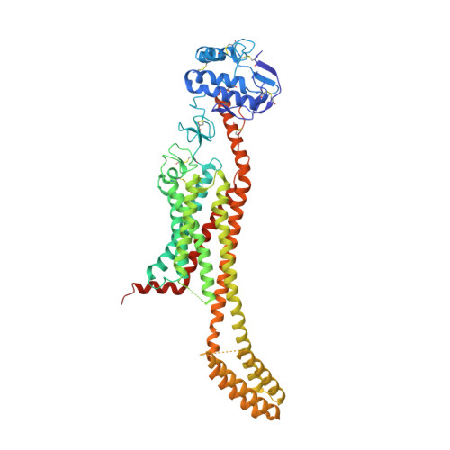
    The seven-transmembrane-spanning protein Smoothened is the central transducer in Hedgehog signaling, a pathway fundamental in development and in cancer. Smoothened is activated by cholesterol binding to its extracellular cysteine-rich domain (CRD). How this interaction leads to changes in the transmembrane domain and Smoothened activation is unknown. Here, we report crystal structures of sterol-activated Smoothened. The CRD undergoes a dramatic reorientation, allosterically causing the transmembrane domain to adopt a conformation similar to active G-protein-coupled receptors. We show that Smoothened contains a unique inhibitory π-cation lock, which is broken on activation and is disrupted in constitutively active oncogenic mutants. Smoothened activation opens a hydrophobic tunnel, suggesting a pathway for cholesterol movement from the inner membrane leaflet to the CRD. All Smoothened antagonists bind the transmembrane domain and block tunnel opening, but cyclopamine also binds the CRD, inducing the active transmembrane conformation. Together, these results define the mechanisms of Smoothened activation and inhibition.


  • Organizational Affiliation

    Department of Cell Biology, Harvard Medical School, 240 Longwood Avenue, Boston, MA 02115, USA.


Macromolecules
Find similar proteins by:  (by identity cutoff)  |  3D Structure
Entity ID: 1
MoleculeChains Sequence LengthOrganismDetailsImage
Smoothened,Soluble cytochrome b562,Smoothened612Xenopus laevisEscherichia coliMutation(s): 4 
Gene Names: smoSmocybC
Membrane Entity: Yes 
UniProt
Find proteins for P0ABE7 (Escherichia coli)
Explore P0ABE7 
Go to UniProtKB:  P0ABE7
Entity Groups  
Sequence Clusters30% Identity50% Identity70% Identity90% Identity95% Identity100% Identity
UniProt GroupP0ABE7
Sequence Annotations
Expand
  • Reference Sequence
Experimental Data & Validation

Experimental Data

  • Method: X-RAY DIFFRACTION
  • Resolution: 3.75 Å
  • R-Value Free: 0.260 
  • R-Value Work: 0.227 
  • R-Value Observed: 0.229 
  • Space Group: C 1 2 1
Unit Cell:
Length ( Å )Angle ( ˚ )
a = 145.778α = 90
b = 79.232β = 108.51
c = 104.076γ = 90
Software Package:
Software NamePurpose
PHENIXrefinement
PDB_EXTRACTdata extraction
DIALSdata reduction
Aimlessdata scaling
PHASERphasing

Structure Validation

View Full Validation Report


Ligand Structure Quality Assessment 


Entry History & Funding Information

Deposition Data


Funding OrganizationLocationGrant Number
National Institutes of Health/National Human Genome Research Institute (NIH/NHGRI)United StatesGM110041

Revision History  (Full details and data files)

  • Version 1.0: 2018-05-23
    Type: Initial release
  • Version 1.1: 2018-06-13
    Changes: Data collection, Database references
  • Version 1.2: 2018-07-25
    Changes: Data collection, Database references
  • Version 1.3: 2019-12-18
    Changes: Author supporting evidence
  • Version 1.4: 2023-10-04
    Changes: Data collection, Database references, Refinement description