6D21

Crystal structure of the FERM domain of zebrafish FARP2

  • Classification: SIGNALING PROTEIN
  • Organism(s): Danio rerio
  • Expression System: Escherichia coli BL21(DE3)
  • Mutation(s): No 

  • Deposited: 2018-04-12 Released: 2018-07-18 
  • Deposition Author(s): Kuo, Y.C., Zhang, X.
  • Funding Organization(s): National Institutes of Health/National Institute of General Medical Sciences (NIH/NIGMS), Welch Foundation, National Natural Science Foundation of China (NSFC)

Experimental Data Snapshot

  • Method: X-RAY DIFFRACTION
  • Resolution: 2.00 Å
  • R-Value Free: 0.239 
  • R-Value Work: 0.183 
  • R-Value Observed: 0.185 

wwPDB Validation   3D Report Full Report


This is version 1.2 of the entry. See complete history


Literature

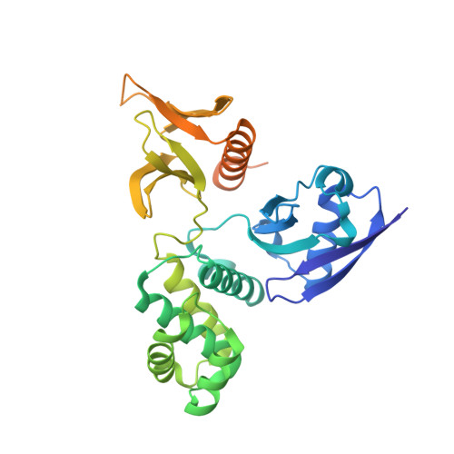
Structural analyses of FERM domain-mediated membrane localization of FARP1.

Kuo, Y.C.He, X.Coleman, A.J.Chen, Y.J.Dasari, P.Liou, J.Biederer, T.Zhang, X.

(2018) Sci Rep 8: 10477-10477

  • DOI: https://doi.org/10.1038/s41598-018-28692-4
  • Primary Citation of Related Structures:  
    6D21, 6D2K, 6D2Q

  • PubMed Abstract: 

    FARP1 is a multi-domain protein that is involved in regulating neuronal development through interacting with cell surface proteins such as class A Plexins and SynCAM 1. The N-terminal FERM domain in FARP1 is known to both promote membrane localization and mediate these protein interactions, for which the underlying molecular mechanisms remain unclear. Here we determined the crystal structures of the FERM domain of FARP1 from zebrafish, and those of FARP2 (a close homolog of FARP1) from mouse and zebrafish. These FERM domains adopt the three-leaved clover fold that is typical of all FERM domains. Our structures reveal a positively charged surface patch that is highly conserved in the FERM domain of FARP1 and FARP2. In vitro lipid-binding experiments showed that the FARP1 FERM domain binds specifically to several types of phospholipid, which is dependent on the positively charged surface patch. We further determined through cell-based analyses that this surface patch on the FERM domain underlies the localization of FARP1 to the plasma membrane, and that FERM domain interactions recruit it to postsynaptic sites in neurons.


  • Organizational Affiliation

    Department of Pharmacology, University of Texas Southwestern Medical Center, Dallas, TX, 75390, USA.


Macromolecules
Find similar proteins by:  (by identity cutoff)  |  3D Structure
Entity ID: 1
MoleculeChains Sequence LengthOrganismDetailsImage
FERM, RhoGEF and pleckstrin domain protein 2333Danio rerioMutation(s): 0 
Gene Names: farp2
UniProt
Find proteins for E7FE33 (Danio rerio)
Explore E7FE33 
Go to UniProtKB:  E7FE33
Entity Groups  
Sequence Clusters30% Identity50% Identity70% Identity90% Identity95% Identity100% Identity
UniProt GroupE7FE33
Sequence Annotations
Expand
  • Reference Sequence
Experimental Data & Validation

Experimental Data

  • Method: X-RAY DIFFRACTION
  • Resolution: 2.00 Å
  • R-Value Free: 0.239 
  • R-Value Work: 0.183 
  • R-Value Observed: 0.185 
  • Space Group: P 21 21 21
Unit Cell:
Length ( Å )Angle ( ˚ )
a = 35.377α = 90
b = 82.723β = 90
c = 96.015γ = 90
Software Package:
Software NamePurpose
PHENIXrefinement
HKL-2000data collection
HKL-2000data scaling
Cootmodel building
PHASERphasing

Structure Validation

View Full Validation Report


Entry History & Funding Information

Deposition Data


Funding OrganizationLocationGrant Number
National Institutes of Health/National Institute of General Medical Sciences (NIH/NIGMS)United StatesGM088197
Welch FoundationUnited StatesI-1702
National Natural Science Foundation of China (NSFC)China#31500605

Revision History  (Full details and data files)

  • Version 1.0: 2018-07-18
    Type: Initial release
  • Version 1.1: 2020-01-01
    Changes: Author supporting evidence
  • Version 1.2: 2024-03-13
    Changes: Data collection, Database references