6CZ8 | pdb_00006cz8

The arsenate respiratory reductase (Arr) complex from Shewanella sp. ANA-3 bound to arsenate


Experimental Data Snapshot

  • Method: X-RAY DIFFRACTION
  • Resolution: 1.78 Å
  • R-Value Free: 
    0.168 (Depositor), 0.163 (DCC) 
  • R-Value Work: 
    0.137 (Depositor), 0.133 (DCC) 
  • R-Value Observed: 
    0.138 (Depositor) 

wwPDB Validation 3D Report Full Report

Validation slider image for 6CZ8

Ligand Structure Quality Assessment 


This is version 1.6 of the entry. See complete history

Literature

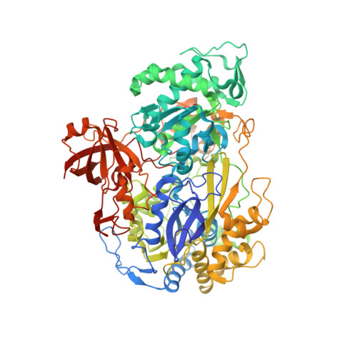
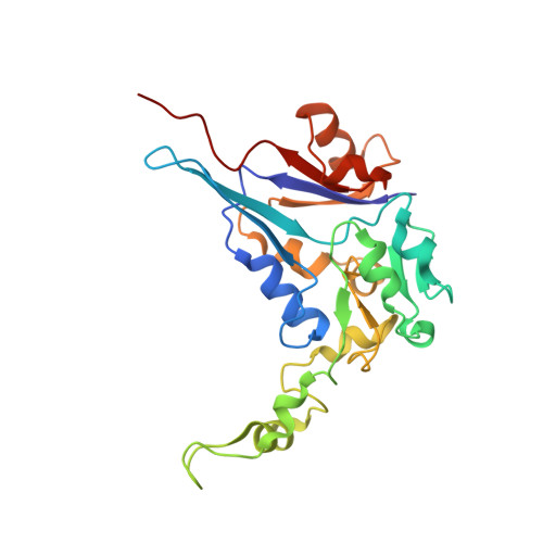
Structural and mechanistic analysis of the arsenate respiratory reductase provides insight into environmental arsenic transformations.

Glasser, N.R.Oyala, P.H.Osborne, T.H.Santini, J.M.Newman, D.K.

(2018) Proc Natl Acad Sci U S A 115: E8614-E8623

  • DOI: https://doi.org/10.1073/pnas.1807984115
  • Primary Citation Related Structures: 
    6CZ7, 6CZ8, 6CZ9, 6CZA

  • PubMed Abstract: 

    Arsenate respiration by bacteria was discovered over two decades ago and is catalyzed by diverse organisms using the well-conserved Arr enzyme complex. Until now, the mechanisms underpinning this metabolism have been relatively opaque. Here, we report the structure of an Arr complex (solved by X-ray crystallography to 1.6-Å resolution), which was enabled by an improved Arr expression method in the genetically tractable arsenate respirer Shewanella sp. ANA-3. We also obtained structures bound with the substrate arsenate (1.8 Å), the product arsenite (1.8 Å), and the natural inhibitor phosphate (1.7 Å). The structures reveal a conserved active-site motif that distinguishes Arr [(R/K)GRY] from the closely related arsenite respiratory oxidase (Arx) complex (XGRGWG). Arr activity assays using methyl viologen as the electron donor and arsenate as the electron acceptor display two-site ping-pong kinetics. A Mo(V) species was detected with EPR spectroscopy, which is typical for proteins with a pyranopterin guanine dinucleotide cofactor. Arr is an extraordinarily fast enzyme that approaches the diffusion limit ( K m = 44.6 ± 1.6 μM, k cat = 9,810 ± 220 seconds -1 ), and phosphate is a competitive inhibitor of arsenate reduction ( K i = 325 ± 12 μM). These observations, combined with knowledge of typical sedimentary arsenate and phosphate concentrations and known rates of arsenate desorption from minerals in the presence of phosphate, suggest that ( i ) arsenate desorption limits microbiologically induced arsenate reductive mobilization and ( ii ) phosphate enhances arsenic mobility by stimulating arsenate desorption rather than by inhibiting it at the enzymatic level.


  • Organizational Affiliation
    • Division of Biology and Biological Engineering, California Institute of Technology, Pasadena, CA 91125.

Macromolecule Content 

  • Total Structure Weight: 241.55 kDa 
  • Atom Count: 19,011 
  • Modeled Residue Count: 2,089 
  • Deposited Residue Count: 2,096 
  • Unique protein chains: 2

Macromolecules

Find similar proteins by:|  3D Structure
Entity ID: 1
MoleculeChains  Sequence LengthOrganismDetailsImage
ArrAA [auth C],
C [auth A]
814Shewanella sp. ANA-3Mutation(s): 0 
Gene Names: arrAShewana3_2341
EC: 1.20.99.1
UniProt
Find proteins for Q7WTU0 (Shewanella sp. (strain ANA-3))
Explore Q7WTU0 
Go to UniProtKB:  Q7WTU0
Entity Groups
Sequence Clusters30% Identity50% Identity70% Identity90% Identity95% Identity100% Identity
UniProt GroupQ7WTU0
Sequence Annotations
Expand
Reference Sequence
Find similar proteins by:|  3D Structure
Entity ID: 2
MoleculeChains  Sequence LengthOrganismDetailsImage
4Fe-4S ferredoxin, iron-sulfur binding domain proteinB [auth D],
D [auth B]
234Shewanella sp. ANA-3Mutation(s): 0 
Gene Names: arrBShewana3_2340
UniProt
Find proteins for Q7WTT9 (Shewanella sp. (strain ANA-3))
Explore Q7WTT9 
Go to UniProtKB:  Q7WTT9
Entity Groups
Sequence Clusters30% Identity50% Identity70% Identity90% Identity95% Identity100% Identity
UniProt GroupQ7WTT9
Sequence Annotations
Expand
Reference Sequence

Small Molecules

Ligands 7 Unique
IDChains Name / Formula / InChI Key2D Diagram3D Interactions
MGD
(Subject of Investigation/LOI)

Query on MGD


Download:Ideal Coordinates CCD File
F [auth C],
G [auth C],
W [auth A],
X [auth A]
2-AMINO-5,6-DIMERCAPTO-7-METHYL-3,7,8A,9-TETRAHYDRO-8-OXA-1,3,9,10-TETRAAZA-ANTHRACEN-4-ONE GUANOSINE DINUCLEOTIDE
C20 H26 N10 O13 P2 S2
VQAGYJCYOLHZDH-ILXWUORBSA-N
SF4

Query on SF4


Download:Ideal Coordinates CCD File
E [auth C]
HA [auth B]
IA [auth B]
JA [auth B]
KA [auth B]
E [auth C],
HA [auth B],
IA [auth B],
JA [auth B],
KA [auth B],
Q [auth D],
R [auth D],
S [auth D],
T [auth D],
V [auth A]
IRON/SULFUR CLUSTER
Fe4 S4
LJBDFODJNLIPKO-UHFFFAOYSA-N
PG5

Query on PG5


Download:Ideal Coordinates CCD File
FA [auth A],
O [auth C]
1-METHOXY-2-[2-(2-METHOXY-ETHOXY]-ETHANE
C8 H18 O4
YFNKIDBQEZZDLK-UHFFFAOYSA-N
ART
(Subject of Investigation/LOI)

Query on ART


Download:Ideal Coordinates CCD File
H [auth C],
Y [auth A]
ARSENATE
As O4
DJHGAFSJWGLOIV-UHFFFAOYSA-K
MO
(Subject of Investigation/LOI)

Query on MO


Download:Ideal Coordinates CCD File
I [auth C],
Z [auth A]
MOLYBDENUM ATOM
Mo
ZOKXTWBITQBERF-UHFFFAOYSA-N
FMT

Query on FMT


Download:Ideal Coordinates CCD File
CA [auth A]
DA [auth A]
EA [auth A]
L [auth C]
M [auth C]
CA [auth A],
DA [auth A],
EA [auth A],
L [auth C],
M [auth C],
N [auth C],
U [auth D]
FORMIC ACID
C H2 O2
BDAGIHXWWSANSR-UHFFFAOYSA-N
O

Query on O


Download:Ideal Coordinates CCD File
AA [auth A]
BA [auth A]
GA [auth A]
J [auth C]
K [auth C]
AA [auth A],
BA [auth A],
GA [auth A],
J [auth C],
K [auth C],
P [auth C]
OXYGEN ATOM
O
XLYOFNOQVPJJNP-UHFFFAOYSA-N

Experimental Data & Validation

Experimental Data

  • Method: X-RAY DIFFRACTION
  • Resolution: 1.78 Å
  • R-Value Free:  0.168 (Depositor), 0.163 (DCC) 
  • R-Value Work:  0.137 (Depositor), 0.133 (DCC) 
  • R-Value Observed: 0.138 (Depositor) 
Space Group: C 1 2 1
Unit Cell:
Length ( Å )Angle ( ˚ )
a = 233.162α = 90
b = 86.344β = 127.67
c = 148.221γ = 90
Software Package:
Software NamePurpose
PHENIXrefinement
XDSdata reduction
XSCALEdata scaling
PHASERphasing

Structure Validation

View Full Validation Report


Ligand Structure Quality Assessment 


Entry History 

& Funding Information

Deposition Data


Funding OrganizationLocationGrant Number
National Institutes of Health/National Heart, Lung, and Blood Institute (NIH/NHLBI)United States5R01HL117328-03

Revision History  (Full details and data files)

  • Version 1.0: 2018-08-15
    Type: Initial release
  • Version 1.1: 2018-08-29
    Changes: Data collection, Database references
  • Version 1.2: 2018-09-19
    Changes: Data collection, Database references
  • Version 1.3: 2018-10-03
    Changes: Data collection, Database references
  • Version 1.4: 2019-02-20
    Changes: Author supporting evidence, Data collection
  • Version 1.5: 2019-12-04
    Changes: Author supporting evidence
  • Version 1.6: 2024-03-13
    Changes: Data collection, Database references, Derived calculations