6CPV | pdb_00006cpv

MicroED structure of NaK ion channel reveals a process of Na+ partition into the selectivity filter


Experimental Data Snapshot

  • Method: ELECTRON CRYSTALLOGRAPHY
  • Resolution: 2.50 Å
  • R-Value Free: 
    0.263 (Depositor) 
  • R-Value Work: 
    0.218 (Depositor) 
  • R-Value Observed: 
    0.221 (Depositor) 

Starting Model: experimental
View more details

wwPDB Validation 3D Report Full Report

Validation slider image for 6CPV

This is version 1.2 of the entry. See complete history

Literature

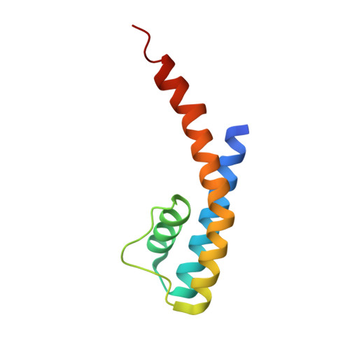
MicroED structure of the NaK ion channel reveals a Na+partition process into the selectivity filter.

Liu, S.Gonen, T.

(2018) Commun Biol 1: 38-38

  • DOI: https://doi.org/10.1038/s42003-018-0040-8
  • Primary Citation Related Structures: 
    6CPV

  • PubMed Abstract: 

    Sodium (Na + ) is a ubiquitous and important inorganic salt mediating many critical biological processes such as neuronal excitation, signaling, and facilitation of various transporters. The hydration states of Na + are proposed to play critical roles in determining the conductance and the selectivity of Na + channels, yet they are rarely captured by conventional structural biology means. Here we use the emerging cryo-electron microscopy (cryoEM) method micro-electron diffraction (MicroED) to study the structure of a prototypical tetrameric Na + -conducting channel, NaK, to 2.5 Å resolution from nano-crystals. Two new conformations at the external site of NaK are identified, allowing us to visualize a partially hydrated Na + ion at the entrance of the channel pore. A process of dilation coupled with Na + movement is identified leading to valuable insights into the mechanism of ion conduction and gating. This study lays the ground work for future studies using MicroED in membrane protein biophysics.


  • Organizational Affiliation
    • Janelia Research Campus, Howard Hughes Medical Institute, 19700 Helix Drive, Ashburn, VA, 20147, USA.

Macromolecule Content 

  • Total Structure Weight: 21.81 kDa 
  • Atom Count: 1,494 
  • Modeled Residue Count: 187 
  • Deposited Residue Count: 192 
  • Unique protein chains: 1

Macromolecules

Find similar proteins by:|  3D Structure
Entity ID: 1
MoleculeChains  Sequence LengthOrganismDetailsImage
Potassium channel protein
A, B
96Bacillus cereusMutation(s): 0 
Gene Names: A9485_19160B4155_3291BACERE00184_02078CN419_22740CN950_06075CN980_22870COI98_17615COK18_26145CON37_12595
Membrane Entity: Yes 
UniProt
Find proteins for Q81HW2 (Bacillus cereus (strain ATCC 14579 / DSM 31 / CCUG 7414 / JCM 2152 / NBRC 15305 / NCIMB 9373 / NCTC 2599 / NRRL B-3711))
Explore Q81HW2 
Go to UniProtKB:  Q81HW2
Entity Groups
Sequence Clusters30% Identity50% Identity70% Identity90% Identity95% Identity100% Identity
UniProt GroupQ81HW2
Sequence Annotations
Expand
Reference Sequence

Small Molecules

Ligands 2 Unique
IDChains Name / Formula / InChI Key2D Diagram3D Interactions
MPD

Query on MPD


Download:Ideal Coordinates CCD File
K [auth A](4S)-2-METHYL-2,4-PENTANEDIOL
C6 H14 O2
SVTBMSDMJJWYQN-YFKPBYRVSA-N
NA

Query on NA


Download:Ideal Coordinates CCD File
C [auth A]
D [auth A]
E [auth A]
F [auth A]
G [auth A]
C [auth A],
D [auth A],
E [auth A],
F [auth A],
G [auth A],
H [auth A],
I [auth A],
J [auth A],
L [auth B],
M [auth B],
N [auth B],
O [auth B]
SODIUM ION
Na
FKNQFGJONOIPTF-UHFFFAOYSA-N

Experimental Data & Validation

Experimental Data

  • Method: ELECTRON CRYSTALLOGRAPHY
  • Resolution: 2.50 Å
  • R-Value Free:  0.263 (Depositor) 
  • R-Value Work:  0.218 (Depositor) 
  • R-Value Observed: 0.221 (Depositor) 
Space Group: I 4
Unit Cell:
Length ( Å )Angle ( ˚ )
a = 68.072α = 90
b = 68.072β = 90
c = 89.3γ = 90
Software Package:
Software NamePurpose
PHENIXrefinement
Aimlessdata scaling
iMOSFLMdata reduction
PHASERphasing

Structure Validation

View Full Validation Report


Entry History 

& Funding Information

Deposition Data


Funding OrganizationLocationGrant Number
Howard Hughes Medical Institute (HHMI)United States1

Revision History  (Full details and data files)

  • Version 1.0: 2018-09-12
    Type: Initial release
  • Version 1.1: 2019-11-20
    Changes: Author supporting evidence
  • Version 1.2: 2023-10-04
    Changes: Data collection, Database references, Derived calculations, Refinement description