6CNA | pdb_00006cna

GluN1-GluN2B NMDA receptors with exon 5


Experimental Data Snapshot

  • Method: ELECTRON MICROSCOPY
  • Resolution: 4.60 Å
  • Aggregation State: PARTICLE 
  • Reconstruction Method: SINGLE PARTICLE 

wwPDB Validation 3D Report Full Report

Validation slider image for 6CNA

This is version 2.2 of the entry. See complete history

Literature

Structural Mechanism of Functional Modulation by Gene Splicing in NMDA Receptors.

Regan, M.C.Grant, T.McDaniel, M.J.Karakas, E.Zhang, J.Traynelis, S.F.Grigorieff, N.Furukawa, H.

(2018) Neuron 98: 521-529.e3

  • DOI: https://doi.org/10.1016/j.neuron.2018.03.034
  • Primary Citation Related Structures: 
    6CNA

  • PubMed Abstract: 

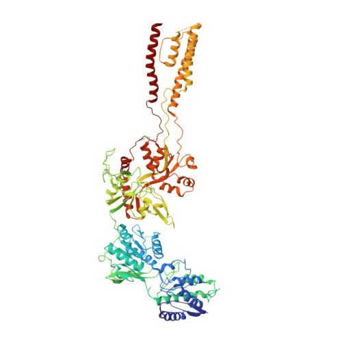
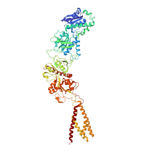
    Alternative gene splicing gives rise to N-methyl-D-aspartate (NMDA) receptor ion channels with defined functional properties and unique contributions to calcium signaling in a given chemical environment in the mammalian brain. Splice variants possessing the exon-5-encoded motif at the amino-terminal domain (ATD) of the GluN1 subunit are known to display robustly altered deactivation rates and pH sensitivity, but the underlying mechanism for this functional modification is largely unknown. Here, we show through cryoelectron microscopy (cryo-EM) that the presence of the exon 5 motif in GluN1 alters the local architecture of heterotetrameric GluN1-GluN2 NMDA receptors and creates contacts with the ligand-binding domains (LBDs) of the GluN1 and GluN2 subunits, which are absent in NMDA receptors lacking the exon 5 motif. The unique interactions established by the exon 5 motif are essential to the stability of the ATD/LBD and LBD/LBD interfaces that are critically involved in controlling proton sensitivity and deactivation.


  • Organizational Affiliation
    • WM Keck Structural Biology Laboratory, Cold Spring Harbor Laboratory, Cold Spring Harbor, NY 11724, USA.

Macromolecule Content 

  • Total Structure Weight: 375.99 kDa 
  • Atom Count: 21,426 
  • Modeled Residue Count: 3,160 
  • Deposited Residue Count: 3,300 
  • Unique protein chains: 2

Macromolecules

Find similar proteins by:|  3D Structure
Entity ID: 1
MoleculeChains  Sequence LengthOrganismDetailsImage
Glutamate receptor ionotropic, NMDA 1
A, C
838Rattus norvegicusMutation(s): 0 
Gene Names: Grin1Nmdar1
Membrane Entity: Yes 
UniProt
Find proteins for P35439 (Rattus norvegicus)
Explore P35439 
Go to UniProtKB:  P35439
Entity Groups
Sequence Clusters30% Identity50% Identity70% Identity90% Identity95% Identity100% Identity
UniProt GroupP35439
Glycosylation
Glycosylation Sites: 4Go to GlyGen: P35439-1
Sequence Annotations
Expand
Reference Sequence
Find similar proteins by:|  3D Structure
Entity ID: 2
MoleculeChains  Sequence LengthOrganismDetailsImage
Glutamate receptor ionotropic, NMDA 2B
B, D
812Rattus norvegicusMutation(s): 0 
Gene Names: Grin2b
Membrane Entity: Yes 
UniProt
Find proteins for Q00960 (Rattus norvegicus)
Explore Q00960 
Go to UniProtKB:  Q00960
Entity Groups
Sequence Clusters30% Identity50% Identity70% Identity90% Identity95% Identity100% Identity
UniProt GroupQ00960
Glycosylation
Glycosylation Sites: 3Go to GlyGen: Q00960-1
Sequence Annotations
Expand
Reference Sequence

Oligosaccharides

Help  
Entity ID: 3
MoleculeChains Length2D Diagram GlycosylationD Interactions
2-acetamido-2-deoxy-beta-D-glucopyranose-(1-4)-2-acetamido-2-deoxy-beta-D-glucopyranose
E, F, G, H, I
E, F, G, H, I, J
2N-Glycosylation
Glycosylation Resources
GlyTouCan: G42666HT
GlyCosmos: G42666HT
GlyGen: G42666HT

Small Molecules

Ligands 1 Unique
IDChains Name / Formula / InChI Key2D Diagram3D Interactions
NAG

Query on NAG


Download:Ideal Coordinates CCD File
K [auth A]
L [auth A]
M [auth A]
N [auth B]
O [auth B]
K [auth A],
L [auth A],
M [auth A],
N [auth B],
O [auth B],
P [auth C],
Q [auth C],
R [auth C],
S [auth D],
T [auth D]
2-acetamido-2-deoxy-beta-D-glucopyranose
C8 H15 N O6
OVRNDRQMDRJTHS-FMDGEEDCSA-N

Experimental Data & Validation

Experimental Data

  • Method: ELECTRON MICROSCOPY
  • Resolution: 4.60 Å
  • Aggregation State: PARTICLE 
  • Reconstruction Method: SINGLE PARTICLE 
EM Software:
TaskSoftware PackageVersion
MODEL REFINEMENTPHENIX

Structure Validation

View Full Validation Report


Entry History 

& Funding Information

Deposition Data


Funding OrganizationLocationGrant Number
National Institutes of Health/National Institute of Mental Health (NIH/NIMH)United StatesMH085926
National Institutes of Health/National Institute of General Medical Sciences (NIH/NIGMS)United StatesGM105730
Howard Hughes Medical Institute (HHMI)United States--

Revision History  (Full details and data files)

  • Version 1.0: 2018-10-03
    Type: Initial release
  • Version 1.1: 2019-11-20
    Changes: Author supporting evidence
  • Version 1.2: 2019-12-18
    Changes: Other
  • Version 2.0: 2020-07-29
    Type: Remediation
    Reason: Carbohydrate remediation
    Changes: Advisory, Atomic model, Data collection, Derived calculations, Structure summary
  • Version 2.1: 2024-10-23
    Changes: Data collection, Database references, Structure summary
  • Version 2.2: 2025-05-14
    Changes: Data collection