Analysis of Global and Site-Specific Radiation Damage in Cryo-EM.
Hattne, J., Shi, D., Glynn, C., Zee, C.T., Gallagher-Jones, M., Martynowycz, M.W., Rodriguez, J.A., Gonen, T.(2018) Structure 26: 759-766.e4
- PubMed: 29706530 Search on PubMedSearch on PubMed Central
- DOI: https://doi.org/10.1016/j.str.2018.03.021
- Primary Citation Related Structures:
6CL7, 6CL8, 6CL9, 6CLA, 6CLB, 6CLC, 6CLD, 6CLE, 6CLF, 6CLG, 6CLH, 6CLI, 6CLJ, 6CLK, 6CLL, 6CLM, 6CLN, 6CLO, 6CLP, 6CLQ, 6CLR, 6CLS, 6CLT - PubMed Abstract:
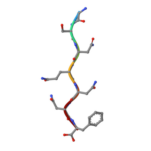
Micro-crystal electron diffraction (MicroED) combines the efficiency of electron scattering with diffraction to allow structure determination from nano-sized crystalline samples in cryoelectron microscopy (cryo-EM). It has been used to solve structures of a diverse set of biomolecules and materials, in some cases to sub-atomic resolution. However, little is known about the damaging effects of the electron beam on samples during such measurements. We assess global and site-specific damage from electron radiation on nanocrystals of proteinase K and of a prion hepta-peptide and find that the dynamics of electron-induced damage follow well-established trends observed in X-ray crystallography. Metal ions are perturbed, disulfide bonds are broken, and acidic side chains are decarboxylated while the diffracted intensities decay exponentially with increasing exposure. A better understanding of radiation damage in MicroED improves our assessment and processing of all types of cryo-EM data.
- Howard Hughes Medical Institute, University of California, Los Angeles, Los Angeles CA 90095, USA; Janelia Research Campus, Howard Hughes Medical Institute, Ashburn, VA 20147, USA.
Organizational Affiliation: