6CK7 | pdb_00006ck7

Crystal structure of a peptide deformylase from Legionella pneumophila bound to actinonin


Experimental Data Snapshot

  • Method: X-RAY DIFFRACTION
  • Resolution: 1.65 Å
  • R-Value Free: 
    0.208 (Depositor), 0.210 (DCC) 
  • R-Value Work: 
    0.182 (Depositor), 0.180 (DCC) 
  • R-Value Observed: 
    0.183 (Depositor) 

Starting Model: experimental
View more details

wwPDB Validation   3D Report Full Report


Ligand Structure Quality Assessment 


This is version 1.2 of the entry. See complete history


Literature

Structures of Legionella pneumophila serogroup 1 peptide deformylase bound to nickel(II) and actinonin.

Nguyen, C.L.Fan, W.Fisher, S.Matthews, K.Norman, J.O.Abendroth, J.Barrett, K.F.Craig, J.K.Edwards, T.E.Lorimer, D.D.McLaughlin, K.J.

(2025) Acta Crystallogr F Struct Biol Commun 81: 163-170

  • DOI: https://doi.org/10.1107/S2053230X25001876
  • Primary Citation of Related Structures:  
    6CK7

  • PubMed Abstract: 

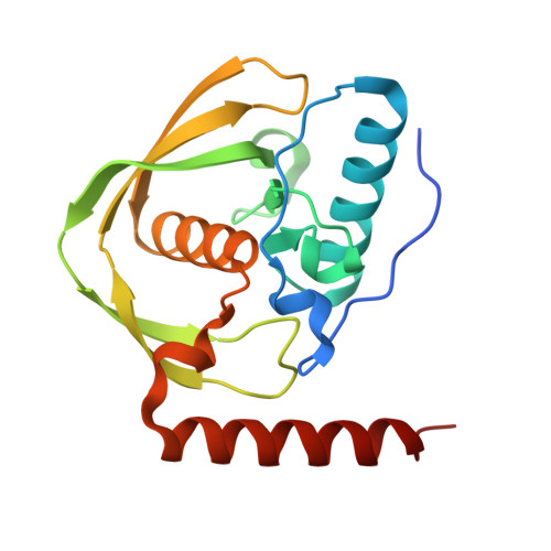
    Legionella pneumophila serogroup 1 is the primary causative agent of Legionnaires' disease, a rare but severe respiratory infection. While the fatality rate of Legionnaires' disease is low in the general population, it is more pronounced in vulnerable communities such as the immunocompromised. Thus, the development of new antimicrobials is of interest for use when existing antibiotics may not be applicable. Peptide deformylases (PDFs) have been under continued investigation as targets for novel antimicrobial compounds. PDF plays an essential role in protein synthesis, removing the N-terminal formyl group from new polypeptides, and is required for growth in most bacteria. Here, we report two crystal structures of L. pneumophila serogroup 1 PDF (LpPDF) bound to either Ni 2+ , an active state, or inhibited by actinonin and Zn 2+ ; the structures were determined to 1.5 and 1.65 Å resolution, respectively, and were solved by the Seattle Structural Genomics Center for Infectious Disease (SSGCID). The SSGCID is charged with determining structures of biologically important proteins and molecules from human pathogens. As actinonin is an antimicrobial natural product that has been used as a reference compound in drug development, these structures will help support the ongoing drug-development process.


  • Organizational Affiliation
    • Biochemistry Program, Vassar College, 124 Raymond Avenue, Poughkeepsie, NY 12604, USA.

Macromolecules
Find similar proteins by:  (by identity cutoff)  |  3D Structure
Entity ID: 1
MoleculeChains Sequence LengthOrganismDetailsImage
Peptide deformylase
A, B, C, D
178Legionella pneumophilaMutation(s): 0 
Gene Names: def_2defdef_1def_3A1D14_13265A9P85_13750ERS240541_01674ERS240560_02189ERS253249_00325lpymg_02824
EC: 3.5.1.88
UniProt
Find proteins for Q5ZSC4 (Legionella pneumophila subsp. pneumophila (strain Philadelphia 1 / ATCC 33152 / DSM 7513))
Explore Q5ZSC4 
Go to UniProtKB:  Q5ZSC4
Entity Groups  
Sequence Clusters30% Identity50% Identity70% Identity90% Identity95% Identity100% Identity
UniProt GroupQ5ZSC4
Sequence Annotations
Expand
  • Reference Sequence
Small Molecules
Ligands 3 Unique
IDChains Name / Formula / InChI Key2D Diagram3D Interactions
BB2
Query on BB2

Download Ideal Coordinates CCD File 
F [auth A],
I [auth B],
L [auth C],
P [auth D]
ACTINONIN
C19 H35 N3 O5
XJLATMLVMSFZBN-VYDXJSESSA-N
SO4
Query on SO4

Download Ideal Coordinates CCD File 
G [auth A],
J [auth B],
M [auth C],
N [auth C],
Q [auth D]
SULFATE ION
O4 S
QAOWNCQODCNURD-UHFFFAOYSA-L
ZN
Query on ZN

Download Ideal Coordinates CCD File 
E [auth A],
H [auth B],
K [auth C],
O [auth D]
ZINC ION
Zn
PTFCDOFLOPIGGS-UHFFFAOYSA-N
Experimental Data & Validation

Experimental Data

  • Method: X-RAY DIFFRACTION
  • Resolution: 1.65 Å
  • R-Value Free:  0.208 (Depositor), 0.210 (DCC) 
  • R-Value Work:  0.182 (Depositor), 0.180 (DCC) 
  • R-Value Observed: 0.183 (Depositor) 
Space Group: P 1 21 1
Unit Cell:
Length ( Å )Angle ( ˚ )
a = 44.65α = 90
b = 132.06β = 103.64
c = 90.38γ = 90
Software Package:
Software NamePurpose
XDSdata reduction
XSCALEdata scaling
PHENIXrefinement
PDB_EXTRACTdata extraction
PHASERphasing

Structure Validation

View Full Validation Report


Ligand Structure Quality Assessment 


Entry History 

Deposition Data

Revision History  (Full details and data files)

  • Version 1.0: 2018-03-07
    Type: Initial release
  • Version 1.1: 2023-10-04
    Changes: Data collection, Database references, Derived calculations, Refinement description
  • Version 1.2: 2025-04-09
    Changes: Database references, Structure summary