6CDR

Human CtBP1 (28-378)

  • Classification: TRANSCRIPTION
  • Organism(s): Homo sapiens
  • Expression System: Escherichia coli
  • Mutation(s): Yes 

  • Deposited: 2018-02-09 Released: 2018-05-09 
  • Deposition Author(s): Royer, W.E., Bellesis, A.G.
  • Funding Organization(s): National Institutes of Health/National Institute of General Medical Sciences (NIH/NIGMS), National Institutes of Health/Office of the Director

Experimental Data Snapshot

  • Method: X-RAY DIFFRACTION
  • Resolution: 2.40 Å
  • R-Value Free: 0.262 
  • R-Value Work: 0.235 
  • R-Value Observed: 0.236 

wwPDB Validation   3D Report Full Report


Ligand Structure Quality Assessment 


This is version 1.4 of the entry. See complete history


Literature

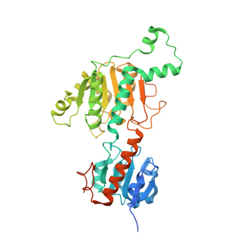
Assembly of human C-terminal binding protein (CtBP) into tetramers.

Bellesis, A.G.Jecrois, A.M.Hayes, J.A.Schiffer, C.A.Royer, W.E.

(2018) J Biol Chem 293: 9101-9112

  • DOI: https://doi.org/10.1074/jbc.RA118.002514
  • Primary Citation of Related Structures:  
    6CDF, 6CDR

  • PubMed Abstract: 

    C-terminal binding protein 1 (CtBP1) and CtBP2 are transcriptional coregulators that repress numerous cellular processes, such as apoptosis, by binding transcription factors and recruiting chromatin-remodeling enzymes to gene promoters. The NAD(H)-linked oligomerization of human CtBP is coupled to its co-transcriptional activity, which is implicated in cancer progression. However, the biologically relevant level of CtBP assembly has not been firmly established; nor has the stereochemical arrangement of the subunits above that of a dimer. Here, multi-angle light scattering (MALS) data established the NAD + - and NADH-dependent assembly of CtBP1 and CtBP2 into tetramers. An examination of subunit interactions within CtBP1 and CtBP2 crystal lattices revealed that both share a very similar tetrameric arrangement resulting from assembly of two dimeric pairs, with specific interactions probably being sensitive to NAD(H) binding. Creating a series of mutants of both CtBP1 and CtBP2, we tested the hypothesis that the crystallographically observed interdimer pairing stabilizes the solution tetramer. MALS data confirmed that these mutants disrupt both CtBP1 and CtBP2 tetramers, with the dimer generally remaining intact, providing the first stereochemical models for tetrameric assemblies of CtBP1 and CtBP2. The crystal structure of a subtle destabilizing mutant suggested that small structural perturbations of the hinge region linking the substrate- and NAD-binding domains are sufficient to weaken the CtBP1 tetramer. These results strongly suggest that the tetramer is important in CtBP function, and the series of CtBP mutants reported here can be used to investigate the physiological role of the tetramer.


  • Organizational Affiliation

    From the Department of Biochemistry and Molecular Pharmacology, University of Massachusetts Medical School, Worcester, Massachusetts 01605 and.


Macromolecules
Find similar proteins by:  (by identity cutoff)  |  3D Structure
Entity ID: 1
MoleculeChains Sequence LengthOrganismDetailsImage
C-terminal-binding protein 1373Homo sapiensMutation(s): 1 
Gene Names: CTBP1CTBP
EC: 1.1.1
UniProt & NIH Common Fund Data Resources
Find proteins for Q13363 (Homo sapiens)
Explore Q13363 
Go to UniProtKB:  Q13363
PHAROS:  Q13363
GTEx:  ENSG00000159692 
Entity Groups  
Sequence Clusters30% Identity50% Identity70% Identity90% Identity95% Identity100% Identity
UniProt GroupQ13363
Sequence Annotations
Expand
  • Reference Sequence
Small Molecules
Ligands 4 Unique
IDChains Name / Formula / InChI Key2D Diagram3D Interactions
NAI
Query on NAI

Download Ideal Coordinates CCD File 
B [auth A]1,4-DIHYDRONICOTINAMIDE ADENINE DINUCLEOTIDE
C21 H29 N7 O14 P2
BOPGDPNILDQYTO-NNYOXOHSSA-N
EDO
Query on EDO

Download Ideal Coordinates CCD File 
E [auth A],
F [auth A],
G [auth A],
H [auth A]
1,2-ETHANEDIOL
C2 H6 O2
LYCAIKOWRPUZTN-UHFFFAOYSA-N
FMT
Query on FMT

Download Ideal Coordinates CCD File 
I [auth A]FORMIC ACID
C H2 O2
BDAGIHXWWSANSR-UHFFFAOYSA-N
CL
Query on CL

Download Ideal Coordinates CCD File 
C [auth A],
D [auth A]
CHLORIDE ION
Cl
VEXZGXHMUGYJMC-UHFFFAOYSA-M
Experimental Data & Validation

Experimental Data

  • Method: X-RAY DIFFRACTION
  • Resolution: 2.40 Å
  • R-Value Free: 0.262 
  • R-Value Work: 0.235 
  • R-Value Observed: 0.236 
  • Space Group: P 64 2 2
Unit Cell:
Length ( Å )Angle ( ˚ )
a = 89.218α = 90
b = 89.218β = 90
c = 164.233γ = 120
Software Package:
Software NamePurpose
PHENIXrefinement
HKL-2000data reduction
HKL-2000data scaling
PHENIXphasing

Structure Validation

View Full Validation Report


Ligand Structure Quality Assessment 


Entry History & Funding Information

Deposition Data


Funding OrganizationLocationGrant Number
National Institutes of Health/National Institute of General Medical Sciences (NIH/NIGMS)United StatesR01 GM119014
National Institutes of Health/Office of the DirectorUnited StatesS10 OD012028

Revision History  (Full details and data files)

  • Version 1.0: 2018-05-09
    Type: Initial release
  • Version 1.1: 2018-06-20
    Changes: Data collection, Database references
  • Version 1.2: 2019-02-20
    Changes: Author supporting evidence, Data collection
  • Version 1.3: 2020-01-01
    Changes: Author supporting evidence
  • Version 1.4: 2023-10-04
    Changes: Data collection, Database references, Refinement description