6C9Q

Mycobacterium tuberculosis adenosine kinase bound to 5'-aminoadenosine


Experimental Data Snapshot

  • Method: X-RAY DIFFRACTION
  • Resolution: 1.95 Å
  • R-Value Free: 0.208 
  • R-Value Work: 0.184 
  • R-Value Observed: 0.185 

wwPDB Validation   3D Report Full Report


This is version 1.3 of the entry. See complete history


Literature

Structure-Guided Drug Design of 6-Substituted Adenosine Analogues as Potent Inhibitors of Mycobacterium tuberculosis Adenosine Kinase.

Crespo, R.A.Dang, Q.Zhou, N.E.Guthrie, L.M.Snavely, T.C.Dong, W.Loesch, K.A.Suzuki, T.You, L.Wang, W.O'Malley, T.Parish, T.Olsen, D.B.Sacchettini, J.C.

(2019) J Med Chem 62: 4483-4499

  • DOI: https://doi.org/10.1021/acs.jmedchem.9b00020
  • Primary Citation of Related Structures:  
    6C67, 6C9N, 6C9P, 6C9Q, 6C9R, 6C9S, 6C9V

  • PubMed Abstract: 

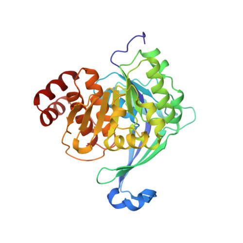
    Mycobacterium tuberculosis adenosine kinase (MtbAdoK) is an essential enzyme of Mtb and forms part of the purine salvage pathway within mycobacteria. Evidence suggests that the purine salvage pathway might play a crucial role in Mtb survival and persistence during its latent phase of infection. In these studies, we adopted a structural approach to the discovery, structure-guided design, and synthesis of a series of adenosine analogues that displayed inhibition constants ranging from 5 to 120 nM against the enzyme. Two of these compounds exhibited low micromolar activity against Mtb with half maximal effective inhibitory concentrations of 1.7 and 4.0 μM. Our selectivity and preliminary pharmacokinetic studies showed that the compounds possess a higher degree of specificity against MtbAdoK when compared with the human counterpart and are well tolerated in rodents, respectively. Finally, crystallographic studies showed the molecular basis of inhibition, potency, and selectivity and revealed the presence of a potentially therapeutically relevant cavity unique to the MtbAdoK homodimer.


  • Organizational Affiliation

    Department of Biochemistry and Biophysics , Texas A&M University , College Station , Texas 77843 , United States.


Macromolecules
Find similar proteins by:  (by identity cutoff)  |  3D Structure
Entity ID: 1
MoleculeChains Sequence LengthOrganismDetailsImage
Adenosine kinase331Mycobacterium tuberculosisMutation(s): 0 
Gene Names: adoKcbhKMRA_2218
EC: 2.7.1.20
UniProt
Find proteins for P9WID5 (Mycobacterium tuberculosis (strain ATCC 25618 / H37Rv))
Explore P9WID5 
Go to UniProtKB:  P9WID5
Entity Groups  
Sequence Clusters30% Identity50% Identity70% Identity90% Identity95% Identity100% Identity
UniProt GroupP9WID5
Sequence Annotations
Expand
  • Reference Sequence
Small Molecules
Ligands 1 Unique
IDChains Name / Formula / InChI Key2D Diagram3D Interactions
5N5
Query on 5N5

Download Ideal Coordinates CCD File 
B [auth A]5'-amino-5'-deoxyadenosine
C10 H14 N6 O3
GVSGUDGNTHCZHI-KQYNXXCUSA-N
Experimental Data & Validation

Experimental Data

  • Method: X-RAY DIFFRACTION
  • Resolution: 1.95 Å
  • R-Value Free: 0.208 
  • R-Value Work: 0.184 
  • R-Value Observed: 0.185 
  • Space Group: P 31 2 1
Unit Cell:
Length ( Å )Angle ( ˚ )
a = 71.904α = 90
b = 71.904β = 90
c = 110.256γ = 120
Software Package:
Software NamePurpose
PHENIXrefinement
HKL-2000data reduction
HKL-2000data scaling
MOLREPphasing

Structure Validation

View Full Validation Report


Entry History & Funding Information

Deposition Data


Funding OrganizationLocationGrant Number
Bill & Melinda Gates FoundationUnited StatesOPP1024055
Welch FoundationUnited StatesA-0015
National Institutes of Health/National Institute Of Allergy and Infectious Diseases (NIH/NIAID)United StatesP01A1095208

Revision History  (Full details and data files)

  • Version 1.0: 2019-05-01
    Type: Initial release
  • Version 1.1: 2019-05-22
    Changes: Data collection, Database references
  • Version 1.2: 2019-12-18
    Changes: Author supporting evidence
  • Version 1.3: 2023-10-04
    Changes: Data collection, Database references, Refinement description