6C96

Cryo-EM structure of mouse TPC1 channel in the apo state

  • Classification: MEMBRANE PROTEIN
  • Organism(s): Mus musculus
  • Expression System: Homo sapiens
  • Mutation(s): No 
  • Membrane Protein: Yes  OPMPDBTMMemProtMDmpstruc

  • Deposited: 2018-01-25 Released: 2018-04-04 
  • Deposition Author(s): She, J., Guo, J., Chen, Q., Bai, X., Jiang, Y.
  • Funding Organization(s): National Institutes of Health/National Center for Research Resources (NIH/NCRR), Welch Foundation, Howard Hughes Medical Institute (HHMI), Cancer Prevention and Research Institute of Texas (CPRIT), Virginia Murchison Linthicum Scholar in Medical Research fund

Experimental Data Snapshot

  • Method: ELECTRON MICROSCOPY
  • Resolution: 3.40 Å
  • Aggregation State: PARTICLE 
  • Reconstruction Method: SINGLE PARTICLE 

wwPDB Validation   3D Report Full Report


This is version 2.0 of the entry. See complete history


Literature

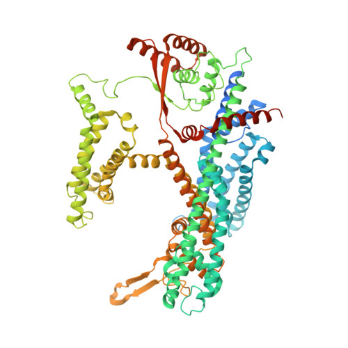
Structural insights into the voltage and phospholipid activation of the mammalian TPC1 channel.

She, J.Guo, J.Chen, Q.Zeng, W.Jiang, Y.Bai, X.C.

(2018) Nature 556: 130-134

  • DOI: https://doi.org/10.1038/nature26139
  • Primary Citation of Related Structures:  
    6C96, 6C9A

  • PubMed Abstract: 

    The organellar two-pore channel (TPC) functions as a homodimer, in which each subunit contains two homologous Shaker-like six-transmembrane (6-TM)-domain repeats. TPCs belong to the voltage-gated ion channel superfamily and are ubiquitously expressed in animals and plants. Mammalian TPC1 and TPC2 are localized at the endolysosomal membrane, and have critical roles in regulating the physiological functions of these acidic organelles. Here we present electron cryo-microscopy structures of mouse TPC1 (MmTPC1)-a voltage-dependent, phosphatidylinositol 3,5-bisphosphate (PtdIns(3,5)P 2 )-activated Na + -selective channel-in both the apo closed state and ligand-bound open state. Combined with functional analysis, these structures provide comprehensive structural insights into the selectivity and gating mechanisms of mammalian TPC channels. The channel has a coin-slot-shaped ion pathway in the filter that defines the selectivity of mammalian TPCs. Only the voltage-sensing domain from the second 6-TM domain confers voltage dependence on MmTPC1. Endolysosome-specific PtdIns(3,5)P 2 binds to the first 6-TM domain and activates the channel under conditions of depolarizing membrane potential. Structural comparisons between the apo and PtdIns(3,5)P 2 -bound structures show the interplay between voltage and ligand in channel activation. These MmTPC1 structures reveal lipid binding and regulation in a 6-TM voltage-gated channel, which is of interest in light of the emerging recognition of the importance of phosphoinositide regulation of ion channels.


  • Organizational Affiliation

    Department of Physiology, University of Texas Southwestern Medical Center, Dallas, Texas 75390-9040, USA.


Macromolecules
Find similar proteins by:  (by identity cutoff)  |  3D Structure
Entity ID: 1
MoleculeChains Sequence LengthOrganismDetailsImage
Two pore calcium channel protein 1
A, B
825Mus musculusMutation(s): 0 
Gene Names: Tpcn1Kiaa1169Tpc1
Membrane Entity: Yes 
UniProt & NIH Common Fund Data Resources
Find proteins for Q9EQJ0 (Mus musculus)
Explore Q9EQJ0 
Go to UniProtKB:  Q9EQJ0
IMPC:  MGI:2182472
Entity Groups  
Sequence Clusters30% Identity50% Identity70% Identity90% Identity95% Identity100% Identity
UniProt GroupQ9EQJ0
Sequence Annotations
Expand
  • Reference Sequence
Oligosaccharides

Help

Entity ID: 2
MoleculeChains Length2D Diagram Glycosylation3D Interactions
2-acetamido-2-deoxy-beta-D-glucopyranose-(1-4)-2-acetamido-2-deoxy-beta-D-glucopyranose
C, D
2N-Glycosylation
Glycosylation Resources
GlyTouCan:  G42666HT
GlyCosmos:  G42666HT
GlyGen:  G42666HT
Experimental Data & Validation

Experimental Data

  • Method: ELECTRON MICROSCOPY
  • Resolution: 3.40 Å
  • Aggregation State: PARTICLE 
  • Reconstruction Method: SINGLE PARTICLE 
EM Software:
TaskSoftware PackageVersion
RECONSTRUCTIONRELION2

Structure Validation

View Full Validation Report


Entry History & Funding Information

Deposition Data


Funding OrganizationLocationGrant Number
National Institutes of Health/National Center for Research Resources (NIH/NCRR)United StatesGM079179
Welch FoundationUnited StatesI-1578
Howard Hughes Medical Institute (HHMI)United States--
Cancer Prevention and Research Institute of Texas (CPRIT)United States--
Virginia Murchison Linthicum Scholar in Medical Research fundUnited States--

Revision History  (Full details and data files)

  • Version 1.0: 2018-04-04
    Type: Initial release
  • Version 1.1: 2018-04-18
    Changes: Data collection, Database references
  • Version 1.2: 2019-04-24
    Changes: Author supporting evidence, Data collection
  • Version 1.3: 2019-11-20
    Changes: Author supporting evidence
  • Version 1.4: 2019-12-18
    Changes: Other
  • Version 2.0: 2020-07-29
    Type: Remediation
    Reason: Carbohydrate remediation
    Changes: Atomic model, Data collection, Derived calculations, Structure summary