6C62

An unexpected vestigial protein complex reveals the evolutionary origins of an s-triazine catabolic enzyme.


Experimental Data Snapshot

  • Method: X-RAY DIFFRACTION
  • Resolution: 1.95 Å
  • R-Value Free: 0.191 
  • R-Value Work: 0.153 
  • R-Value Observed: 0.155 

wwPDB Validation   3D Report Full Report


This is version 1.1 of the entry. See complete history


Literature

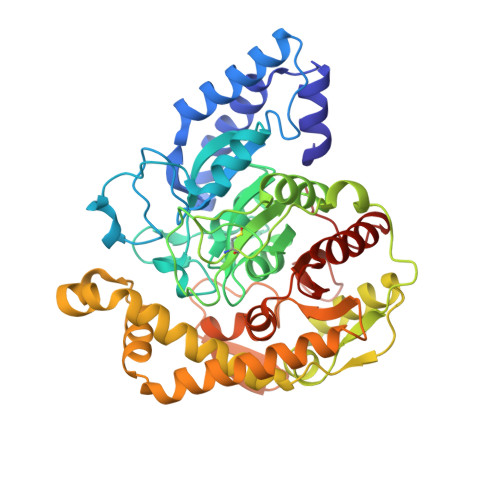
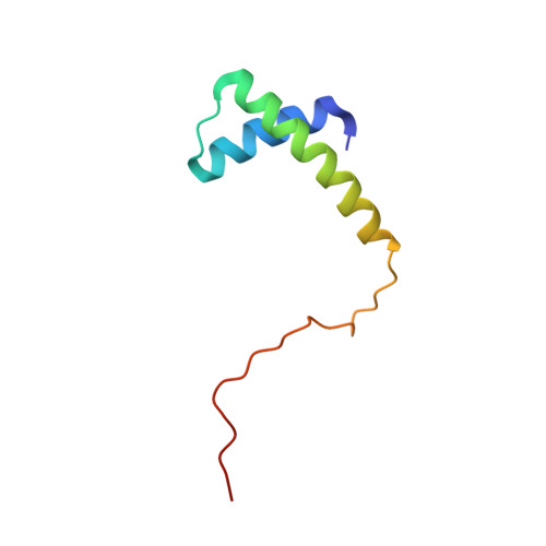
An unexpected vestigial protein complex reveals the evolutionary origins of ans-triazine catabolic enzyme.

Esquirol, L.Peat, T.S.Wilding, M.Liu, J.W.French, N.G.Hartley, C.J.Onagi, H.Nebl, T.Easton, C.J.Newman, J.Scott, C.

(2018) J Biol Chem 293: 7880-7891

  • DOI: https://doi.org/10.1074/jbc.RA118.001996
  • Primary Citation of Related Structures:  
    6C62, 6C6G

  • PubMed Abstract: 
  • Cyanuric acid is a metabolic intermediate of s -triazines, such as atrazine (a common herbicide) and melamine (used in resins and plastics). Cyanuric acid is mineralized to ammonia and carbon dioxide by the soil bacterium Pseudomonas sp. strain ADP via three hydrolytic enzymes (AtzD, AtzE, and AtzF) ...

    Cyanuric acid is a metabolic intermediate of s -triazines, such as atrazine (a common herbicide) and melamine (used in resins and plastics). Cyanuric acid is mineralized to ammonia and carbon dioxide by the soil bacterium Pseudomonas sp. strain ADP via three hydrolytic enzymes (AtzD, AtzE, and AtzF). Here, we report the purification and biochemical and structural characterization of AtzE. Contrary to previous reports, we found that AtzE is not a biuret amidohydrolase, but instead it catalyzes the hydrolytic deamination of 1-carboxybiuret. X-ray crystal structures of apo AtzE and AtzE bound with the suicide inhibitor phenyl phosphorodiamidate revealed that the AtzE enzyme complex consists of two independent molecules in the asymmetric unit. We also show that AtzE forms an α2β2 heterotetramer with a previously unidentified 68-amino acid-long protein (AtzG) encoded in the cyanuric acid mineralization operon from Pseudomonas sp. strain ADP. Moreover, we observed that AtzG is essential for the production of soluble, active AtzE and that this obligate interaction is a vestige of their shared evolutionary origin. We propose that AtzEG was likely recruited into the cyanuric acid-mineralizing pathway from an ancestral glutamine transamidosome that required protein-protein interactions to enforce the exclusion of solvent from the transamidation reaction.


    Organizational Affiliation

    Synthetic Biology Future Science Platform, CSIRO Land and Water, Canberra, Australian Capital Territory 2601.


Macromolecules
Find similar proteins by:  (by identity cutoff)  |  3D Structure
Entity ID: 1
MoleculeChainsSequence LengthOrganismDetailsImage
Biuret hydrolase
A, B
457Pseudomonas sp. ADPMutation(s): 0 
Gene Names: atzE
EC: 3.5.1.84 (PDB Primary Data), 3.5.1.131 (UniProt)
UniProt
Find proteins for Q936X3 (Pseudomonas sp. (strain ADP))
Explore Q936X3 
Go to UniProtKB:  Q936X3
Entity Groups  
Sequence Clusters30% Identity50% Identity70% Identity90% Identity95% Identity100% Identity
UniProt GroupQ936X3
Protein Feature View
Expand
  • Reference Sequence
Find similar proteins by:  (by identity cutoff)  |  3D Structure
Entity ID: 2
MoleculeChainsSequence LengthOrganismDetailsImage
AtzG
C, D
68Pseudomonas sp. ADPMutation(s): 0 
Gene Names: atzG
UniProt
Find proteins for A0A384E126 (Pseudomonas sp. (strain ADP))
Explore A0A384E126 
Go to UniProtKB:  A0A384E126
Entity Groups  
Sequence Clusters30% Identity50% Identity70% Identity90% Identity95% Identity100% Identity
UniProt GroupA0A384E126
Protein Feature View
Expand
  • Reference Sequence
Small Molecules
Ligands 1 Unique
IDChainsName / Formula / InChI Key2D Diagram3D Interactions
MG
Query on MG

Download Ideal Coordinates CCD File 
E [auth B],
F [auth D],
G [auth D]
MAGNESIUM ION
Mg
JLVVSXFLKOJNIY-UHFFFAOYSA-N
 Ligand Interaction
Modified Residues  1 Unique
IDChainsTypeFormula2D DiagramParent
CSO
Query on CSO
A, B
L-PEPTIDE LINKINGC3 H7 N O3 SCYS
Experimental Data & Validation

Experimental Data

  • Method: X-RAY DIFFRACTION
  • Resolution: 1.95 Å
  • R-Value Free: 0.191 
  • R-Value Work: 0.153 
  • R-Value Observed: 0.155 
  • Space Group: I 1 2 1
Unit Cell:
Length ( Å )Angle ( ˚ )
a = 79.489α = 90
b = 89.04β = 101.89
c = 141.672γ = 90
Software Package:
Software NamePurpose
REFMACrefinement
DIALSdata reduction
Aimlessdata scaling
MoRDaphasing

Structure Validation

View Full Validation Report


Entry History 

Deposition Data

Revision History  (Full details and data files)

  • Version 1.0: 2018-03-21
    Type: Initial release
  • Version 1.1: 2018-05-30
    Changes: Data collection, Database references, Source and taxonomy, Structure summary