6C4R

Staphylopine dehydrogenase (SaODH) - Apo


Experimental Data Snapshot

  • Method: X-RAY DIFFRACTION
  • Resolution: 2.29 Å
  • R-Value Free: 0.268 
  • R-Value Work: 0.207 
  • R-Value Observed: 0.212 

wwPDB Validation   3D Report Full Report


This is version 1.4 of the entry. See complete history


Literature

Staphylopine, pseudopaline, and yersinopine dehydrogenases: A structural and kinetic analysis of a new functional class of opine dehydrogenase.

McFarlane, J.S.Davis, C.L.Lamb, A.L.

(2018) J Biol Chem 293: 8009-8019

  • DOI: https://doi.org/10.1074/jbc.RA118.002007
  • Primary Citation of Related Structures:  
    6C4L, 6C4M, 6C4N, 6C4R, 6C4T

  • PubMed Abstract: 

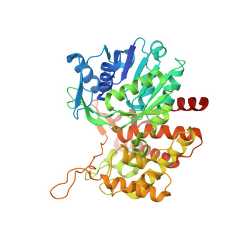
    Opine dehydrogenases (ODHs) from the bacterial pathogens Staphylococcus aureus , Pseudomonas aeruginosa , and Yersinia pestis perform the final enzymatic step in the biosynthesis of a new class of opine metallophores, which includes staphylopine, pseudopaline, and yersinopine, respectively. Growing evidence indicates an important role for this pathway in metal acquisition and virulence, including in lung and burn-wound infections ( P. aeruginosa ) and in blood and heart infections ( S. aureus ). Here, we present kinetic and structural characterizations of these three opine dehydrogenases. A steady-state kinetic analysis revealed that the three enzymes differ in α-keto acid and NAD(P)H substrate specificity and nicotianamine-like substrate stereoselectivity. The structural basis for these differences was determined from five ODH X-ray crystal structures, ranging in resolution from 1.9 to 2.5 Å, with or without NADP + bound. Variation in hydrogen bonding with NADPH suggested an explanation for the differential recognition of this substrate by these three enzymes. Our analysis further revealed candidate residues in the active sites required for binding of the α-keto acid and nicotianamine-like substrates and for catalysis. This work reports the first structural kinetic analyses of enzymes involved in opine metallophore biosynthesis in three important bacterial pathogens of humans.


  • Organizational Affiliation

    Department of Molecular Biosciences, University of Kansas, Lawrence, Kansas 66045.


Macromolecules
Find similar proteins by:  (by identity cutoff)  |  3D Structure
Entity ID: 1
MoleculeChains Sequence LengthOrganismDetailsImage
Staphylopine dehydrogenase450Staphylococcus aureusMutation(s): 0 
Gene Names: AB466_12420AB526_12775AFP37_00945APW47_05390
UniProt
Find proteins for A0A0H3JT80 (Staphylococcus aureus (strain Mu50 / ATCC 700699))
Explore A0A0H3JT80 
Go to UniProtKB:  A0A0H3JT80
Entity Groups  
Sequence Clusters30% Identity50% Identity70% Identity90% Identity95% Identity100% Identity
UniProt GroupA0A0H3JT80
Sequence Annotations
Expand
  • Reference Sequence
Experimental Data & Validation

Experimental Data

  • Method: X-RAY DIFFRACTION
  • Resolution: 2.29 Å
  • R-Value Free: 0.268 
  • R-Value Work: 0.207 
  • R-Value Observed: 0.212 
  • Space Group: P 21 21 2
Unit Cell:
Length ( Å )Angle ( ˚ )
a = 187.741α = 90
b = 49.092β = 90
c = 58.585γ = 90
Software Package:
Software NamePurpose
PHENIXrefinement
XDSdata reduction
XSCALEdata scaling
PHASERphasing

Structure Validation

View Full Validation Report


Entry History & Funding Information

Deposition Data


Funding OrganizationLocationGrant Number
National Science Foundation (NSF, United States)United StatesCHE1403293
National Institutes of Health/National Institute of General Medical Sciences (NIH/NIGMS)United StatesT32GM008545
National Institutes of Health/National Institute of General Medical Sciences (NIH/NIGMS)United StatesP20GM103418

Revision History  (Full details and data files)

  • Version 1.0: 2018-04-11
    Type: Initial release
  • Version 1.1: 2018-04-25
    Changes: Data collection, Database references
  • Version 1.2: 2018-06-06
    Changes: Data collection, Database references
  • Version 1.3: 2019-11-27
    Changes: Author supporting evidence
  • Version 1.4: 2023-10-04
    Changes: Data collection, Database references, Refinement description