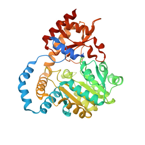
6C3D

O2-, PLP-dependent L-arginine hydroxylase RohP quinonoid II complex


Experimental Data Snapshot

  • Method: X-RAY DIFFRACTION
  • Resolution: 1.55 Å
  • R-Value Free: 0.186 
  • R-Value Work: 0.160 
  • R-Value Observed: 0.161 

Starting Model: experimental
View more details

wwPDB Validation   3D Report Full Report


Ligand Structure Quality Assessment 


This is version 1.5 of the entry. See complete history


Literature

Snapshots of the Catalytic Cycle of an O2, Pyridoxal Phosphate-Dependent Hydroxylase.

Hedges, J.B.Kuatsjah, E.Du, Y.L.Eltis, L.D.Ryan, K.S.

(2018) ACS Chem Biol 13: 965-974

  • DOI: https://doi.org/10.1021/acschembio.8b00039
  • Primary Citation of Related Structures:  
    6C3A, 6C3B, 6C3C, 6C3D

  • PubMed Abstract: 

    Enzymes that catalyze hydroxylation of unactivated carbons normally contain heme and nonheme iron cofactors. By contrast, how a pyridoxal phosphate (PLP)-dependent enzyme could catalyze such a hydroxylation was unknown. Here, we investigate RohP, a PLP-dependent enzyme that converts l-arginine to ( S)-4-hydroxy-2-ketoarginine. We determine that the RohP reaction consumes oxygen with stoichiometric release of H 2 O 2 . To understand this unusual chemistry, we obtain ∼1.5 Å resolution structures that capture intermediates along the catalytic cycle. Our data suggest that RohP carries out a four-electron oxidation and a stereospecific alkene hydration to give the ( S)-configured product. Together with our earlier studies on an O 2 , PLP-dependent l-arginine oxidase, our work suggests that there is a shared pathway leading to both oxidized and hydroxylated products from l-arginine.


  • Organizational Affiliation

    Institute of Pharmaceutical Biotechnology, School of Medicine , Zhejiang University , Hangzhou , China.


Macromolecules
Find similar proteins by:  (by identity cutoff)  |  3D Structure
Entity ID: 1
MoleculeChains Sequence LengthOrganismDetailsImage
Uncharacterized protein
A, B
413Streptantibioticus cattleyicolor NRRL 8057 = DSM 46488Mutation(s): 0 
Gene Names: SCATT_03970
UniProt
Find proteins for G8WNK6 (Streptantibioticus cattleyicolor (strain ATCC 35852 / DSM 46488 / JCM 4925 / NBRC 14057 / NRRL 8057))
Explore G8WNK6 
Go to UniProtKB:  G8WNK6
Entity Groups  
Sequence Clusters30% Identity50% Identity70% Identity90% Identity95% Identity100% Identity
UniProt GroupG8WNK6
Sequence Annotations
Expand
  • Reference Sequence
Small Molecules
Ligands 3 Unique
IDChains Name / Formula / InChI Key2D Diagram3D Interactions
EMS
Query on EMS

Download Ideal Coordinates CCD File 
I [auth A],
Q [auth B]
(2E,3E)-5-carbamimidamido-2-{[(Z)-{3-hydroxy-2-methyl-5-[(phosphonooxy)methyl]pyridin-4(1H)-ylidene}methyl]imino}pent-3-enoic acid
C14 H20 N5 O7 P
DYVCEYAQAUKHHQ-PUVXEYBGSA-N
PEG
Query on PEG

Download Ideal Coordinates CCD File 
P [auth B]DI(HYDROXYETHYL)ETHER
C4 H10 O3
MTHSVFCYNBDYFN-UHFFFAOYSA-N
EDO
Query on EDO

Download Ideal Coordinates CCD File 
C [auth A]
D [auth A]
E [auth A]
F [auth A]
G [auth A]
C [auth A],
D [auth A],
E [auth A],
F [auth A],
G [auth A],
H [auth A],
J [auth B],
K [auth B],
L [auth B],
M [auth B],
N [auth B],
O [auth B]
1,2-ETHANEDIOL
C2 H6 O2
LYCAIKOWRPUZTN-UHFFFAOYSA-N
Experimental Data & Validation

Experimental Data

  • Method: X-RAY DIFFRACTION
  • Resolution: 1.55 Å
  • R-Value Free: 0.186 
  • R-Value Work: 0.160 
  • R-Value Observed: 0.161 
  • Space Group: C 1 2 1
Unit Cell:
Length ( Å )Angle ( ˚ )
a = 79.01α = 90
b = 112.56β = 96.26
c = 102.13γ = 90
Software Package:
Software NamePurpose
PHENIXrefinement
Aimlessdata scaling
PDB_EXTRACTdata extraction
iMOSFLMdata reduction
PHASERphasing

Structure Validation

View Full Validation Report


Ligand Structure Quality Assessment 


Entry History & Funding Information

Deposition Data


Funding OrganizationLocationGrant Number
Natural Sciences and Engineering Research Council (NSERC, Canada)CanadaRGPIN-2016-03778
Alfred P. Sloan FoundationUnited StatesFG-20166503

Revision History  (Full details and data files)

  • Version 1.0: 2018-03-07
    Type: Initial release
  • Version 1.1: 2018-04-18
    Changes: Data collection, Derived calculations, Structure summary
  • Version 1.2: 2018-05-02
    Changes: Data collection, Database references, Derived calculations, Structure summary
  • Version 1.3: 2019-02-20
    Changes: Author supporting evidence, Data collection
  • Version 1.4: 2020-01-08
    Changes: Author supporting evidence
  • Version 1.5: 2023-10-04
    Changes: Data collection, Database references, Refinement description