


Clinical signature variant of HCV NS3/4A protease uses a novel mechanism to confer resistance
Matthew, A.N., Schiffer, C.A.To be published.
Experimental Data Snapshot
Starting Model: experimental
View more details
Entity ID: 1 | |||||
|---|---|---|---|---|---|
| Molecule | Chains | Sequence Length | Organism | Details | Image |
| NS4A COFACTOR-NS3 PROTEIN CHIMERA | 203 | hepatitis C virus genotype 1a | Mutation(s): 17 | 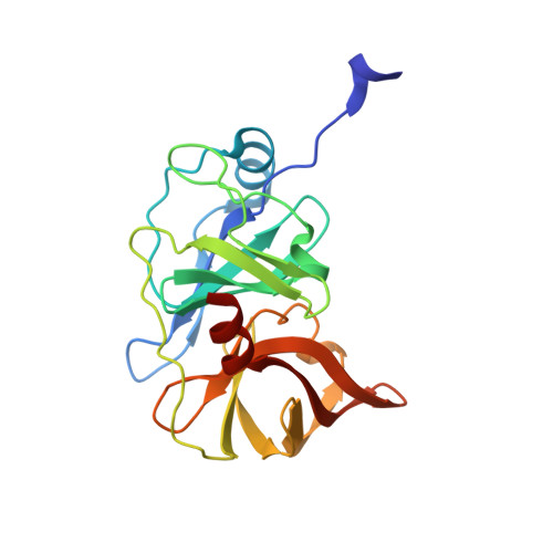 | |
UniProt | |||||
Entity Groups | |||||
| Sequence Clusters | 30% Identity50% Identity70% Identity90% Identity95% Identity100% Identity | ||||
| UniProt Group | A8DG50 | ||||
Sequence AnnotationsExpand | |||||
Reference Sequence | |||||
| Ligands 3 Unique | |||||
|---|---|---|---|---|---|
| ID | Chains | Name / Formula / InChI Key | 2D Diagram | 3D Interactions | |
| TSV Download:Ideal Coordinates CCD File | D [auth A] | (2R,6S,12Z,13aS,14aR,16aS)-6-[(tert-butoxycarbonyl)amino]-14a-[(cyclopropylsulfonyl)carbamoyl]-5,16-dioxo-1,2,3,5,6,7,8
,9,10,11,13a,14,14a,15,16,16a-hexadecahydrocyclopropa[e]pyrrolo[1,2-a][1,4]diazacyclopentadecin-2-yl 4-fluoro-2H-isoindole-2-carboxylate C35 H44 F N5 O9 S YUWURHBDLJOUAP-JSZLBQEHSA-N |  | ||
| SO4 Download:Ideal Coordinates CCD File | C [auth A] | SULFATE ION O4 S QAOWNCQODCNURD-UHFFFAOYSA-L |  | ||
| ZN Download:Ideal Coordinates CCD File | B [auth A] | ZINC ION Zn PTFCDOFLOPIGGS-UHFFFAOYSA-N |  | ||
| Length ( Å ) | Angle ( ˚ ) |
|---|---|
| a = 59.964 | α = 90 |
| b = 55.355 | β = 90 |
| c = 58.891 | γ = 90 |
| Software Name | Purpose |
|---|---|
| HKL-3000 | data scaling |
| PHASER | phasing |
| PHENIX | refinement |
| Coot | model building |
| HKL-3000 | data collection |
| HKL-3000 | data processing |
| Funding Organization | Location | Grant Number |
|---|---|---|
| National Institutes of Health/National Institute Of Allergy and Infectious Diseases (NIH/NIAID) | United States | R01 AI085051 |
| National Institutes of Health/National Institute of General Medical Sciences (NIH/NIGMS) | United States | F31 GM119345 |


