6BZP

STGGYG from low-complexity domain of FUS, residues 77-82


Experimental Data Snapshot

  • Method: ELECTRON CRYSTALLOGRAPHY
  • Resolution: 1.10 Å
  • R-Value Free: 0.255 
  • R-Value Work: 0.219 
  • R-Value Observed: 0.223 

wwPDB Validation   3D Report Full Report


This is version 1.5 of the entry. See complete history


Literature

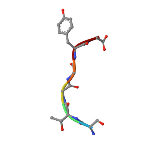
Atomic structures of low-complexity protein segments reveal kinked beta sheets that assemble networks.

Hughes, M.P.Sawaya, M.R.Boyer, D.R.Goldschmidt, L.Rodriguez, J.A.Cascio, D.Chong, L.Gonen, T.Eisenberg, D.S.

(2018) Science 359: 698-701

  • DOI: https://doi.org/10.1126/science.aan6398
  • Primary Citation of Related Structures:  
    6BWZ, 6BXV, 6BXX, 6BZM, 6BZP

  • PubMed Abstract: 

    Subcellular membraneless assemblies are a reinvigorated area of study in biology, with spirited scientific discussions on the forces between the low-complexity protein domains within these assemblies. To illuminate these forces, we determined the atomic structures of five segments from protein low-complexity domains associated with membraneless assemblies. Their common structural feature is the stacking of segments into kinked β sheets that pair into protofilaments. Unlike steric zippers of amyloid fibrils, the kinked sheets interact weakly through polar atoms and aromatic side chains. By computationally threading the human proteome on our kinked structures, we identified hundreds of low-complexity segments potentially capable of forming such interactions. These segments are found in proteins as diverse as RNA binders, nuclear pore proteins, and keratins, which are known to form networks and localize to membraneless assemblies.


  • Organizational Affiliation

    Department of Biological Chemistry and Department of Chemistry and Biochemistry, University of California Los Angeles (UCLA), Howard Hughes Medical Institute (HHMI), UCLA-Department of Energy (DOE) Institute for Genomics and Proteomics, Los Angeles, CA 90095, USA.


Macromolecules

Find similar proteins by:  Sequence   |   3D Structure  

Entity ID: 1
MoleculeChains Sequence LengthOrganismDetailsImage
RNA-binding protein FUS
A, B
6Homo sapiensMutation(s): 0 
UniProt & NIH Common Fund Data Resources
Find proteins for P35637 (Homo sapiens)
Explore P35637 
Go to UniProtKB:  P35637
PHAROS:  P35637
GTEx:  ENSG00000089280 
Entity Groups  
UniProt GroupP35637
Sequence Annotations
Expand
  • Reference Sequence
Small Molecules
Ligands 1 Unique
IDChains Name / Formula / InChI Key2D Diagram3D Interactions
TOE
Query on TOE

Download Ideal Coordinates CCD File 
C [auth B]2-[2-(2-METHOXY-ETHOXY)-ETHOXY]-ETHOXYL
C7 H16 O4
JLGLQAWTXXGVEM-UHFFFAOYSA-N
Experimental Data & Validation

Experimental Data

  • Method: ELECTRON CRYSTALLOGRAPHY
  • Resolution: 1.10 Å
  • R-Value Free: 0.255 
  • R-Value Work: 0.219 
  • R-Value Observed: 0.223 
  • Space Group: P 21 21 21
Unit Cell:
Length ( Å )Angle ( ˚ )
a = 13.79α = 90
b = 4.93β = 90
c = 101.9γ = 90
Software Package:
Software NamePurpose
XSCALEdata scaling
BUSTERrefinement
PDB_EXTRACTdata extraction
EM Software:
TaskSoftware PackageVersion
RECONSTRUCTIONSHELXD2013/2
MODEL REFINEMENTBUSTER2.10.3

Structure Validation

View Full Validation Report


Entry History & Funding Information

Deposition Data


Funding OrganizationLocationGrant Number
Howard Hughes Medical Institute (HHMI)United States--
National Institutes of Health/Office of the DirectorUnited StatesAG-04182
National Science Foundation (NSF, United States)United StatesMCB-0958111

Revision History  (Full details and data files)

  • Version 1.0: 2018-04-04
    Type: Initial release
  • Version 1.1: 2018-04-25
    Changes: Data collection
  • Version 1.2: 2019-11-06
    Changes: Author supporting evidence, Data collection
  • Version 1.3: 2019-11-20
    Changes: Author supporting evidence
  • Version 1.4: 2021-06-30
    Changes: Data collection
  • Version 1.5: 2024-03-13
    Changes: Data collection, Database references