6BXZ

Crystal Structure of Pig Protocadherin-15 EC10-MAD12


Experimental Data Snapshot

  • Method: X-RAY DIFFRACTION
  • Resolution: 2.09 Å
  • R-Value Free: 0.217 
  • R-Value Work: 0.187 
  • R-Value Observed: 0.189 

wwPDB Validation   3D Report Full Report


This is version 1.4 of the entry. See complete history


Literature

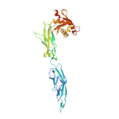
A Mechanically Weak Extracellular Membrane-Adjacent Domain Induces Dimerization of Protocadherin-15.

De-la-Torre, P.Choudhary, D.Araya-Secchi, R.Narui, Y.Sotomayor, M.

(2018) Biophys J 115: 2368-2385

  • DOI: https://doi.org/10.1016/j.bpj.2018.11.010
  • Primary Citation of Related Structures:  
    6BXZ

  • PubMed Abstract: 

    The cadherin superfamily of proteins is defined by the presence of extracellular cadherin (EC) "repeats" that engage in protein-protein interactions to mediate cell-cell adhesion, cell signaling, and mechanotransduction. The extracellular domains of nonclassical cadherins often have a large number of EC repeats along with other subdomains of various folds. Protocadherin-15 (PCDH15), a protein component of the inner-ear tip link filament essential for mechanotransduction, has 11 EC repeats and a membrane adjacent domain (MAD12) of atypical fold. Here we report the crystal structure of a pig PCDH15 fragment including EC10, EC11, and MAD12 in a parallel dimeric arrangement. MAD12 has a unique molecular architecture and folds as a ferredoxin-like domain similar to that found in the nucleoporin protein Nup54. Analytical ultracentrifugation experiments along with size-exclusion chromatography coupled to multiangle laser light scattering and small-angle x-ray scattering corroborate the crystallographic dimer and show that MAD12 induces parallel dimerization of PCDH15 near its membrane insertion point. In addition, steered molecular dynamics simulations suggest that MAD12 is mechanically weak and may unfold before tip-link rupture. Sequence analyses and structural modeling predict the existence of similar domains in cadherin-23, protocadherin-24, and the "giant" FAT and CELSR cadherins, indicating that some of them may also exhibit MAD-induced parallel dimerization.


  • Organizational Affiliation

    Department of Chemistry and Biochemistry, The Ohio State University, Columbus, Ohio.


Macromolecules
Find similar proteins by:  (by identity cutoff)  |  3D Structure
Entity ID: 1
MoleculeChains Sequence LengthOrganismDetailsImage
Protocadherin related 15A [auth C],
B [auth A]
356Sus scrofaMutation(s): 0 
Gene Names: PCDH15
UniProt
Find proteins for F1SD06 (Sus scrofa)
Explore F1SD06 
Go to UniProtKB:  F1SD06
Entity Groups  
Sequence Clusters30% Identity50% Identity70% Identity90% Identity95% Identity100% Identity
UniProt GroupF1SD06
Sequence Annotations
Expand
  • Reference Sequence
Experimental Data & Validation

Experimental Data

  • Method: X-RAY DIFFRACTION
  • Resolution: 2.09 Å
  • R-Value Free: 0.217 
  • R-Value Work: 0.187 
  • R-Value Observed: 0.189 
  • Space Group: P 1 21 1
Unit Cell:
Length ( Å )Angle ( ˚ )
a = 92.284α = 90
b = 49.246β = 92.15
c = 108.253γ = 90
Software Package:
Software NamePurpose
REFMACrefinement
HKL-2000data reduction
HKL-2000data scaling
PHASERphasing

Structure Validation

View Full Validation Report


Entry History & Funding Information

Deposition Data


Funding OrganizationLocationGrant Number
National Institutes of Health/National Institute on Deafness and Other Communication Disorders (NIH/NIDCD)United StatesR01 DC015271
National Institutes of Health/National Institute on Deafness and Other Communication Disorders (NIH/NIDCD)United StatesR00 DC012534

Revision History  (Full details and data files)

  • Version 1.0: 2018-11-28
    Type: Initial release
  • Version 1.1: 2018-12-26
    Changes: Data collection, Database references
  • Version 1.2: 2019-01-02
    Changes: Data collection, Database references
  • Version 1.3: 2019-12-11
    Changes: Author supporting evidence
  • Version 1.4: 2023-10-04
    Changes: Data collection, Database references, Derived calculations, Refinement description