6BWM

Crystal structure of the TRPV2 ion channel


Experimental Data Snapshot

  • Method: X-RAY DIFFRACTION
  • Resolution: 3.90 Å
  • R-Value Free: 0.293 
  • R-Value Work: 0.244 
  • R-Value Observed: 0.247 

wwPDB Validation   3D Report Full Report


This is version 1.4 of the entry. See complete history


Literature

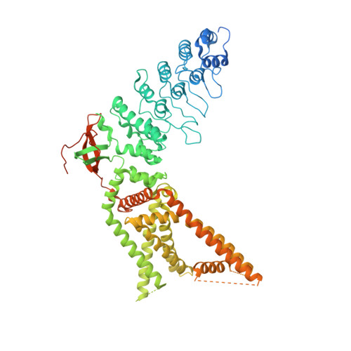
Conformational plasticity in the selectivity filter of the TRPV2 ion channel.

Zubcevic, L.Le, S.Yang, H.Lee, S.Y.

(2018) Nat Struct Mol Biol 25: 405-415

  • DOI: https://doi.org/10.1038/s41594-018-0059-z
  • Primary Citation of Related Structures:  
    6BWJ, 6BWM

  • PubMed Abstract: 

    Transient receptor potential vanilloid (TRPV) channels are activated by ligands and heat and are involved in various physiological processes. In contrast to the architecturally related voltage-gated cation channels, TRPV1 and TRPV2 subtypes possess another activation gate at the selectivity filter that can open widely enough to permeate large organic cations. Despite recent structural advances, the mechanism of selectivity filter gating and permeation for both metal ions and large molecules by TRPV1 or TRPV2 is not well known. Here, we determined two crystal structures of rabbit TRPV2 in its Ca 2+ -bound and resiniferatoxin (RTx)- and Ca 2+ -bound forms, to 3.9 Å and 3.1 Å, respectively. Notably, our structures show that RTx binding leads to two-fold symmetric opening of the selectivity filter of TRPV2 that is wide enough for large organic cation permeation. Combined with functional characterizations, our studies reveal a structural basis for permeation of Ca 2+ and large organic cations in TRPV2.


  • Organizational Affiliation

    Department of Biochemistry, Duke University School of Medicine, Durham, NC, USA.


Macromolecules
Find similar proteins by:  (by identity cutoff)  |  3D Structure
Entity ID: 1
MoleculeChains Sequence LengthOrganismDetailsImage
Transient receptor potential cation channel subfamily V member 2
A, B
776Oryctolagus cuniculusMutation(s): 0 
Gene Names: TRPV2
Membrane Entity: Yes 
UniProt
Find proteins for G1SNM3 (Oryctolagus cuniculus)
Explore G1SNM3 
Go to UniProtKB:  G1SNM3
Entity Groups  
Sequence Clusters30% Identity50% Identity70% Identity90% Identity95% Identity100% Identity
UniProt GroupG1SNM3
Sequence Annotations
Expand
  • Reference Sequence
Small Molecules
Ligands 1 Unique
IDChains Name / Formula / InChI Key2D Diagram3D Interactions
CA
Query on CA

Download Ideal Coordinates CCD File 
C [auth B]CALCIUM ION
Ca
BHPQYMZQTOCNFJ-UHFFFAOYSA-N
Experimental Data & Validation

Experimental Data

  • Method: X-RAY DIFFRACTION
  • Resolution: 3.90 Å
  • R-Value Free: 0.293 
  • R-Value Work: 0.244 
  • R-Value Observed: 0.247 
  • Space Group: P 2 21 21
Unit Cell:
Length ( Å )Angle ( ˚ )
a = 92.028α = 90
b = 122.356β = 90
c = 187.209γ = 90
Software Package:
Software NamePurpose
PHENIXrefinement
XDSdata reduction
Aimlessdata scaling
PHASERphasing

Structure Validation

View Full Validation Report


Entry History & Funding Information

Deposition Data


Funding OrganizationLocationGrant Number
National Institutes of Health/National Institute of Neurological Disorders and Stroke (NIH/NINDS)United States1R35NS097241

Revision History  (Full details and data files)

  • Version 1.0: 2018-05-02
    Type: Initial release
  • Version 1.1: 2018-05-23
    Changes: Data collection, Database references
  • Version 1.2: 2019-02-20
    Changes: Author supporting evidence, Data collection
  • Version 1.3: 2019-12-18
    Changes: Author supporting evidence
  • Version 1.4: 2023-10-04
    Changes: Data collection, Database references, Refinement description