Biological Evaluation and X-ray Co-crystal Structures of Cyclohexylpyrrolidine Ligands for Trypanothione Reductase, an Enzyme from the Redox Metabolism of Trypanosoma.
De Gasparo, R., Brodbeck-Persch, E., Bryson, S., Hentzen, N.B., Kaiser, M., Pai, E.F., Krauth-Siegel, R.L., Diederich, F.(2018) ChemMedChem 13: 957-967
- PubMed: 29624890 Search on PubMed
- DOI: https://doi.org/10.1002/cmdc.201800067
- Primary Citation Related Structures:
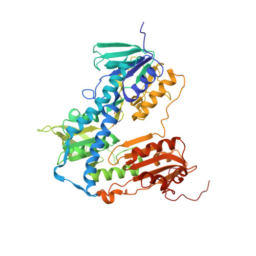
6BTL, 6BU7 - PubMed Abstract:
The tropical diseases human African trypanosomiasis, Chagas disease, and the various forms of leishmaniasis are caused by parasites of the family of trypanosomatids. These protozoa possess a unique redox metabolism based on trypanothione and trypanothione reductase (TR), making TR a promising drug target. We report the optimization of properties and potency of cyclohexylpyrrolidine inhibitors of TR by structure-based design. The best inhibitors were freely soluble and showed competitive inhibition constants (K i ) against Trypanosoma (T.) brucei TR and T. cruzi TR and in vitro activities (half-maximal inhibitory concentration, IC 50 ) against these parasites in the low micromolar range, with high selectivity against human glutathione reductase. X-ray co-crystal structures confirmed the binding of the ligands to the hydrophobic wall of the "mepacrine binding site" with the new, solubility-providing vectors oriented toward the surface of the large active site.
- Laboratorium für Organische Chemie, ETH Zürich, Vladimir-Prelog-Weg 3, 8093, Zürich, Switzerland.
Organizational Affiliation: