6BML

Structure of human DHHC20 palmitoyltransferase, irreversibly inhibited by 2-bromopalmitate


Experimental Data Snapshot

  • Method: X-RAY DIFFRACTION
  • Resolution: 2.95 Å
  • R-Value Free: 0.245 
  • R-Value Work: 0.236 
  • R-Value Observed: 0.237 

wwPDB Validation   3D Report Full Report


Ligand Structure Quality Assessment 


This is version 1.2 of the entry. See complete history


Literature

Fatty acyl recognition and transfer by an integral membraneS-acyltransferase.

Rana, M.S.Kumar, P.Lee, C.J.Verardi, R.Rajashankar, K.R.Banerjee, A.

(2018) Science 359

  • DOI: https://doi.org/10.1126/science.aao6326
  • Primary Citation of Related Structures:  
    6BML, 6BMM, 6BMN, 6BMS

  • PubMed Abstract: 

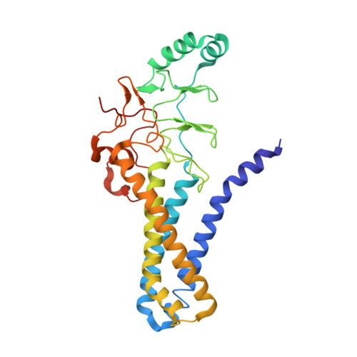
    DHHC (Asp-His-His-Cys) palmitoyltransferases are eukaryotic integral membrane enzymes that catalyze protein palmitoylation, which is important in a range of physiological processes, including small guanosine triphosphatase (GTPase) signaling, cell adhesion, and neuronal receptor scaffolding. We present crystal structures of two DHHC palmitoyltransferases and a covalent intermediate mimic. The active site resides at the membrane-cytosol interface, which allows the enzyme to catalyze thioester-exchange chemistry by using fatty acyl-coenzyme A and explains why membrane-proximal cysteines are candidates for palmitoylation. The acyl chain binds in a cavity formed by the transmembrane domain. We propose a mechanism for acyl chain-length selectivity in DHHC enzymes on the basis of cavity mutants with preferences for shorter and longer acyl chains.


  • Organizational Affiliation

    Cell Biology and Neurobiology Branch, National Institutes of Child Health and Human Development, National Institutes of Health, Bethesda, MD 20892, USA.


Macromolecules
Find similar proteins by:  (by identity cutoff)  |  3D Structure
Entity ID: 1
MoleculeChains Sequence LengthOrganismDetailsImage
human DHHC20 palmitoyltransferase
A, B
295Homo sapiensMutation(s): 0 
Gene Names: ZDHHC20
EC: 2.3.1.225
Membrane Entity: Yes 
UniProt & NIH Common Fund Data Resources
Find proteins for Q5W0Z9 (Homo sapiens)
Explore Q5W0Z9 
Go to UniProtKB:  Q5W0Z9
PHAROS:  Q5W0Z9
GTEx:  ENSG00000180776 
Entity Groups  
Sequence Clusters30% Identity50% Identity70% Identity90% Identity95% Identity100% Identity
UniProt GroupQ5W0Z9
Sequence Annotations
Expand
  • Reference Sequence
Small Molecules
Ligands 4 Unique
IDChains Name / Formula / InChI Key2D Diagram3D Interactions
PAP
Query on PAP

Download Ideal Coordinates CCD File 
J [auth B]3'-PHOSPHATE-ADENOSINE-5'-DIPHOSPHATE
C10 H16 N5 O13 P3
GBBWIZKLHXYJOA-KQYNXXCUSA-N
PLM
Query on PLM

Download Ideal Coordinates CCD File 
E [auth A],
I [auth B]
PALMITIC ACID
C16 H32 O2
IPCSVZSSVZVIGE-UHFFFAOYSA-N
PO4
Query on PO4

Download Ideal Coordinates CCD File 
F [auth A],
K [auth B]
PHOSPHATE ION
O4 P
NBIIXXVUZAFLBC-UHFFFAOYSA-K
ZN
Query on ZN

Download Ideal Coordinates CCD File 
C [auth A],
D [auth A],
G [auth B],
H [auth B]
ZINC ION
Zn
PTFCDOFLOPIGGS-UHFFFAOYSA-N
Experimental Data & Validation

Experimental Data

  • Method: X-RAY DIFFRACTION
  • Resolution: 2.95 Å
  • R-Value Free: 0.245 
  • R-Value Work: 0.236 
  • R-Value Observed: 0.237 
  • Space Group: P 63
Unit Cell:
Length ( Å )Angle ( ˚ )
a = 99.794α = 90
b = 99.794β = 90
c = 159.947γ = 120
Software Package:
Software NamePurpose
PHENIXrefinement
Aimlessdata scaling
PHASERphasing
PDB_EXTRACTdata extraction
Blu-Icedata collection
XDSdata reduction

Structure Validation

View Full Validation Report


Ligand Structure Quality Assessment 


Entry History 

Deposition Data

Revision History  (Full details and data files)

  • Version 1.0: 2018-01-24
    Type: Initial release
  • Version 1.1: 2018-02-21
    Changes: Database references
  • Version 1.2: 2018-03-28
    Changes: Data collection, Database references