6BGP

Crystal Structure of Human Calpain-3 Protease Core Mutant-C129A


Experimental Data Snapshot

  • Method: X-RAY DIFFRACTION
  • Resolution: 2.75 Å
  • R-Value Free: 0.258 
  • R-Value Work: 0.209 
  • R-Value Observed: 0.212 

wwPDB Validation   3D Report Full Report


This is version 1.4 of the entry. See complete history


Literature

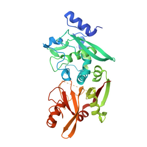
Structures of human calpain-3 protease core with and without bound inhibitor reveal mechanisms of calpain activation.

Ye, Q.Campbell, R.L.Davies, P.L.

(2018) J Biol Chem 293: 4056-4070

  • DOI: https://doi.org/10.1074/jbc.RA117.001097
  • Primary Citation of Related Structures:  
    6BDT, 6BGP, 6BJD, 6BKJ

  • PubMed Abstract: 

    Limb-girdle muscular dystrophy type 2a arises from mutations in the Ca 2+ -activated intracellular cysteine protease calpain-3. This calpain isoform is abundant in skeletal muscle and differs from the main isoforms, calpain-1 and -2, in being a homodimer and having two short insertion sequences. The first of these, IS1, interrupts the protease core and must be cleaved for activation and substrate binding. Here, to learn how calpain-3 can be regulated and inhibited, we determined the structures of the calpain-3 protease core with IS1 present or proteolytically excised. To prevent intramolecular IS1 autoproteolysis, we converted the active-site Cys to Ala. Small-angle X-ray scattering (SAXS) analysis of the C129A mutant suggested that IS1 is disordered and mobile enough to occupy several locations. Surprisingly, this was also true for the apo version of this mutant. We therefore concluded that IS1 might have a binding partner in the sarcomere and is unstructured in its absence. After autoproteolytic IS1 removal from the active Cys 129 calpain-3 protease core, we could solve its crystal structures with and without the cysteine protease inhibitors E-64 and leupeptin covalently bound to the active-site cysteine. In each structure, the active state of the protease core was assembled by the cooperative binding of two Ca 2+ ions to the equivalent sites used in calpain-1 and -2. These structures of the calpain-3 active site with residual IS1 and with bound E-64 and leupeptin may help guide the design of calpain-3-specific inhibitors.


  • Organizational Affiliation

    From the Department of Biomedical and Molecular Sciences, Queen's University, Kingston, Ontario K7L 3N6, Canada.


Macromolecules
Find similar proteins by:  (by identity cutoff)  |  3D Structure
Entity ID: 1
MoleculeChains Sequence LengthOrganismDetailsImage
Calpain-3
A, B, C, D
382Homo sapiensMutation(s): 1 
Gene Names: CAPN3CANP3CANPL3NCL1
EC: 3.4.22.54
UniProt & NIH Common Fund Data Resources
Find proteins for P20807 (Homo sapiens)
Explore P20807 
Go to UniProtKB:  P20807
PHAROS:  P20807
GTEx:  ENSG00000092529 
Entity Groups  
Sequence Clusters30% Identity50% Identity70% Identity90% Identity95% Identity100% Identity
UniProt GroupP20807
Sequence Annotations
Expand
  • Reference Sequence
Small Molecules
Ligands 2 Unique
IDChains Name / Formula / InChI Key2D Diagram3D Interactions
CA
Query on CA

Download Ideal Coordinates CCD File 
E [auth A]
F [auth A]
H [auth B]
I [auth B]
K [auth C]
E [auth A],
F [auth A],
H [auth B],
I [auth B],
K [auth C],
L [auth C],
N [auth D],
O [auth D]
CALCIUM ION
Ca
BHPQYMZQTOCNFJ-UHFFFAOYSA-N
CL
Query on CL

Download Ideal Coordinates CCD File 
G [auth A],
J [auth B],
M [auth C],
P [auth D]
CHLORIDE ION
Cl
VEXZGXHMUGYJMC-UHFFFAOYSA-M
Experimental Data & Validation

Experimental Data

  • Method: X-RAY DIFFRACTION
  • Resolution: 2.75 Å
  • R-Value Free: 0.258 
  • R-Value Work: 0.209 
  • R-Value Observed: 0.212 
  • Space Group: P 21 21 21
Unit Cell:
Length ( Å )Angle ( ˚ )
a = 60.1α = 90
b = 106.18β = 90
c = 225.62γ = 90
Software Package:
Software NamePurpose
REFMACrefinement
PDB_EXTRACTdata extraction
XDSdata reduction
XSCALEdata scaling
PHASERphasing

Structure Validation

View Full Validation Report


Entry History & Funding Information

Deposition Data


Funding OrganizationLocationGrant Number
Canadian Institutes of Health Research (CIHR)CanadaMOP 74681

Revision History  (Full details and data files)

  • Version 1.0: 2018-02-07
    Type: Initial release
  • Version 1.1: 2018-02-14
    Changes: Database references
  • Version 1.2: 2018-05-02
    Changes: Data collection, Database references
  • Version 1.3: 2020-01-08
    Changes: Author supporting evidence
  • Version 1.4: 2023-10-04
    Changes: Data collection, Database references, Refinement description