6BBL

Crystal structure of the a-96Gln MoFe protein variant in the presence of the substrate acetylene


Experimental Data Snapshot

  • Method: X-RAY DIFFRACTION
  • Resolution: 1.68 Å
  • R-Value Free: 0.185 
  • R-Value Work: 0.151 
  • R-Value Observed: 0.153 

wwPDB Validation   3D Report Full Report


Ligand Structure Quality Assessment 


This is version 1.4 of the entry. See complete history


Literature

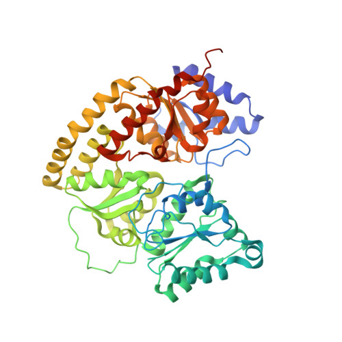
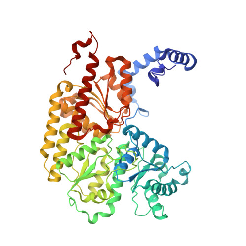
Structural characterization of the nitrogenase molybdenum-iron protein with the substrate acetylene trapped near the active site.

Keable, S.M.Vertemara, J.Zadvornyy, O.A.Eilers, B.J.Danyal, K.Rasmussen, A.J.De Gioia, L.Zampella, G.Seefeldt, L.C.Peters, J.W.

(2017) J Inorg Biochem 180: 129-134

  • DOI: https://doi.org/10.1016/j.jinorgbio.2017.12.008
  • Primary Citation of Related Structures:  
    6BBL

  • PubMed Abstract: 

    The biological reduction of dinitrogen (N 2 ) to ammonia is catalyzed by the complex metalloenzyme nitrogenase. Structures of the nitrogenase component proteins, Iron (Fe) protein and Molybdenum‑iron (MoFe) protein, and the stabilized complexes these component proteins, have been determined, providing a foundation for a number of fundamental aspects of the complicated catalytic mechanism. The reduction of dinitrogen to ammonia is a complex process that involves the binding of N 2 followed by reduction with multiple electrons and protons. Electron transfer into nitrogenase is typically constrained to the unique electron donor, the Fe protein. These constraints have prevented structural characterization of the active site with bound substrate. Recently it has been realized that selected amino acid substitutions in the environment of the active site metal cluster (Iron‑molybdenum cofactor, FeMo-co) allow substrates to persist even in the resting state. Reported here is a 1.70Å crystal structure of a nitrogenase MoFe protein α-96 Arg➔Gln variant with the alternative substrate acetylene trapped in a channel in close proximity to FeMo-co. Complementary theoretical calculations support the validity of the acetylene interaction at this site and is also consistent with more favorable interactions in the variant MoFe protein compared to the native MoFe protein. This work represents the first structural evidence of a substrate trapped in the nitrogenase MoFe protein and is consistent with earlier assignments of proposed substrate pathways and substrate binding sites deduced from biochemical, spectroscopic, and theoretical studies.


  • Organizational Affiliation

    Department of Chemistry and Biochemistry, Montana State University, Bozeman, MT 59717, United States.


Macromolecules
Find similar proteins by:  (by identity cutoff)  |  3D Structure
Entity ID: 1
MoleculeChains Sequence LengthOrganismDetailsImage
Nitrogenase molybdenum-iron protein alpha chain
A, C
492Azotobacter vinelandiiMutation(s): 1 
EC: 1.18.6.1
UniProt
Find proteins for P07328 (Azotobacter vinelandii)
Explore P07328 
Go to UniProtKB:  P07328
Entity Groups  
Sequence Clusters30% Identity50% Identity70% Identity90% Identity95% Identity100% Identity
UniProt GroupP07328
Sequence Annotations
Expand
  • Reference Sequence
Find similar proteins by:  (by identity cutoff)  |  3D Structure
Entity ID: 2
MoleculeChains Sequence LengthOrganismDetailsImage
Nitrogenase molybdenum-iron protein beta chain
B, D
523Azotobacter vinelandiiMutation(s): 0 
EC: 1.18.6.1
UniProt
Find proteins for P07329 (Azotobacter vinelandii)
Explore P07329 
Go to UniProtKB:  P07329
Entity Groups  
Sequence Clusters30% Identity50% Identity70% Identity90% Identity95% Identity100% Identity
UniProt GroupP07329
Sequence Annotations
Expand
  • Reference Sequence
Small Molecules
Ligands 7 Unique
IDChains Name / Formula / InChI Key2D Diagram3D Interactions
ICS
Query on ICS

Download Ideal Coordinates CCD File 
H [auth A],
O [auth C]
iron-sulfur-molybdenum cluster with interstitial carbon
C Fe7 Mo S9
DDQFAOMIVKLFON-UHFFFAOYSA-N
1CL
Query on 1CL

Download Ideal Coordinates CCD File 
I [auth A],
Q [auth C]
FE(8)-S(7) CLUSTER, OXIDIZED
Fe8 S7
JKVMXLBGZBULKV-UHFFFAOYSA-N
HCA
Query on HCA

Download Ideal Coordinates CCD File 
G [auth A],
N [auth C]
3-HYDROXY-3-CARBOXY-ADIPIC ACID
C7 H10 O7
XKJVEVRQMLKSMO-SSDOTTSWSA-N
IMD
Query on IMD

Download Ideal Coordinates CCD File 
F [auth A],
P [auth C],
R [auth D]
IMIDAZOLE
C3 H5 N2
RAXXELZNTBOGNW-UHFFFAOYSA-O
FE
Query on FE

Download Ideal Coordinates CCD File 
J [auth B],
L [auth B]
FE (III) ION
Fe
VTLYFUHAOXGGBS-UHFFFAOYSA-N
C2H
Query on C2H

Download Ideal Coordinates CCD File 
E [auth A],
M [auth C]
acetylene
C2 H2
HSFWRNGVRCDJHI-UHFFFAOYSA-N
MG
Query on MG

Download Ideal Coordinates CCD File 
K [auth B]MAGNESIUM ION
Mg
JLVVSXFLKOJNIY-UHFFFAOYSA-N
Experimental Data & Validation

Experimental Data

  • Method: X-RAY DIFFRACTION
  • Resolution: 1.68 Å
  • R-Value Free: 0.185 
  • R-Value Work: 0.151 
  • R-Value Observed: 0.153 
  • Space Group: P 1 21 1
Unit Cell:
Length ( Å )Angle ( ˚ )
a = 76.781α = 90
b = 128.251β = 109.11
c = 107.272γ = 90
Software Package:
Software NamePurpose
PHENIXrefinement
XDSdata reduction
HKL-2000data reduction
XSCALEdata scaling
SCALEPACKdata scaling
PHENIXphasing

Structure Validation

View Full Validation Report


Ligand Structure Quality Assessment 


Entry History & Funding Information

Deposition Data


Funding OrganizationLocationGrant Number
National Science Foundation (NSF, United States)United StatesMCB-1330807
Department of Energy (DOE, United States)United StatesDE-AC02-76SF00515

Revision History  (Full details and data files)

  • Version 1.0: 2018-01-10
    Type: Initial release
  • Version 1.1: 2018-01-17
    Changes: Author supporting evidence
  • Version 1.2: 2018-08-22
    Changes: Data collection, Database references, Source and taxonomy, Structure summary
  • Version 1.3: 2019-11-27
    Changes: Author supporting evidence
  • Version 1.4: 2023-10-04
    Changes: Data collection, Database references, Derived calculations, Refinement description