Exceptionally tight membrane-binding may explain the key role of the synaptotagmin-7 C2A domain in asynchronous neurotransmitter release.
Voleti, R., Tomchick, D.R., Sudhof, T.C., Rizo, J.(2017) Proc Natl Acad Sci U S A 114: E8518-E8527
- PubMed: 28923929 Search on PubMedSearch on PubMed Central
- DOI: https://doi.org/10.1073/pnas.1710708114
- Primary Citation Related Structures:
6ANJ, 6ANK - PubMed Abstract:
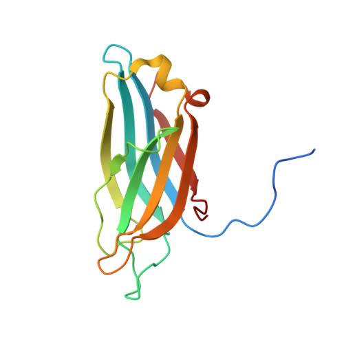
Synaptotagmins (Syts) act as Ca 2+ sensors in neurotransmitter release by virtue of Ca 2+ -binding to their two C 2 domains, but their mechanisms of action remain unclear. Puzzlingly, Ca 2+ -binding to the C 2 B domain appears to dominate Syt1 function in synchronous release, whereas Ca 2+ -binding to the C 2 A domain mediates Syt7 function in asynchronous release. Here we show that crystal structures of the Syt7 C 2 A domain and C 2 AB region, and analyses of intrinsic Ca 2+ -binding to the Syt7 C2 domains using isothermal titration calorimetry, did not reveal major differences that could explain functional differentiation between Syt7 and Syt1. However, using liposome titrations under Ca 2+ saturating conditions, we show that the Syt7 C 2 A domain has a very high membrane affinity and dominates phospholipid binding to Syt7 in the presence or absence of l-α-phosphatidylinositol 4,5-diphosphate (PIP 2 ). For Syt1, the two Ca 2+ -saturated C 2 domains have similar affinities for membranes lacking PIP 2 , but the C 2 B domain dominates binding to PIP 2 -containing membranes. Mutagenesis revealed that the dramatic differences in membrane affinity between the Syt1 and Syt7 C 2 A domains arise in part from apparently conservative residue substitutions, showing how striking biochemical and functional differences can result from the cumulative effects of subtle residue substitutions. Viewed together, our results suggest that membrane affinity may be a key determinant of the functions of Syt C 2 domains in neurotransmitter release.
- Department of Biophysics, University of Texas Southwestern Medical Center, Dallas, TX 75390.
Organizational Affiliation: