6AGK | pdb_00006agk

The structure of CH-II-77-tubulin complex


Experimental Data Snapshot

  • Method: X-RAY DIFFRACTION
  • Resolution: 2.80 Å
  • R-Value Free: 
    0.264 (Depositor), 0.261 (DCC) 
  • R-Value Work: 
    0.211 (Depositor), 0.213 (DCC) 
  • R-Value Observed: 
    0.214 (Depositor) 

Starting Model: experimental
View more details

wwPDB Validation 3D Report Full Report

Validation slider image for 6AGK

Ligand Structure Quality Assessment 


This is version 1.2 of the entry. See complete history

Literature

Structure-Activity Relationship Study of Novel 6-Aryl-2-benzoyl-pyridines as Tubulin Polymerization Inhibitors with Potent Antiproliferative Properties.

Chen, H.Deng, S.Wang, Y.Albadari, N.Kumar, G.Ma, D.Li, W.White, S.W.Miller, D.D.Li, W.

(2020) J Med Chem 63: 827-846

  • DOI: https://doi.org/10.1021/acs.jmedchem.9b01815
  • Primary Citation Related Structures: 
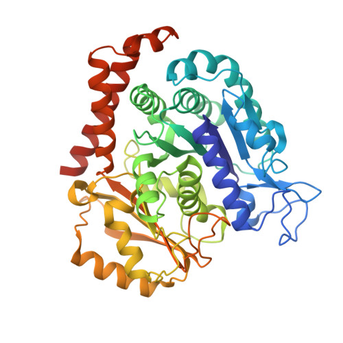
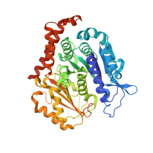
    6AGK, 6PC4

  • PubMed Abstract: 

    We recently reported the crystal structure of tubulin in complex with a colchicine binding site inhibitor (CBSI), ABI-231, having 2-aryl-4-benzoyl-imidazole (ABI). Based on this and additional crystal structures, here we report the structure-activity relationship study of a novel series of pyridine analogues of ABI-231, with compound 4v being the most potent one (average IC 50 ∼ 1.8 nM) against a panel of cancer cell lines. We determined the crystal structures of another potent CBSI ABI-274 and 4v in complex with tubulin and confirmed their direct binding at the colchicine site. 4v inhibited tubulin polymerization, strongly suppressed A375 melanoma tumor growth, induced tumor necrosis, disrupted tumor angiogenesis, and led to tumor cell apoptosis in vivo. Collectively, these studies suggest that 4v represents a promising new generation of tubulin inhibitors.


  • Organizational Affiliation
    • Department of Pharmaceutical Sciences, College of Pharmacy , University of Tennessee Health Science Center , Memphis , Tennessee 38163 , United States.

Macromolecule Content 

  • Total Structure Weight: 265.17 kDa 
  • Atom Count: 17,398 
  • Modeled Residue Count: 2,151 
  • Deposited Residue Count: 2,317 
  • Unique protein chains: 4

Macromolecules

Find similar proteins by:|  3D Structure
Entity ID: 1
MoleculeChains  Sequence LengthOrganismDetailsImage
Tubulin alpha-1B chain
A, C
450Sus scrofaMutation(s): 0 
Gene Names: TUBA1B
EC: 3.6.5
UniProt
Find proteins for Q2XVP4 (Sus scrofa)
Explore Q2XVP4 
Go to UniProtKB:  Q2XVP4
Entity Groups
Sequence Clusters30% Identity50% Identity70% Identity90% Identity95% Identity100% Identity
UniProt GroupQ2XVP4
Sequence Annotations
Expand
Reference Sequence
Find similar proteins by:|  3D Structure
Entity ID: 2
MoleculeChains  Sequence LengthOrganismDetailsImage
Tubulin beta-2B chain
B, D
445Bos taurusMutation(s): 0 
Gene Names: TUBB2B
UniProt
Find proteins for Q6B856 (Bos taurus)
Explore Q6B856 
Go to UniProtKB:  Q6B856
Entity Groups
Sequence Clusters30% Identity50% Identity70% Identity90% Identity95% Identity100% Identity
UniProt GroupQ6B856
Sequence Annotations
Expand
Reference Sequence
Find similar proteins by:|  3D Structure
Entity ID: 3
MoleculeChains  Sequence LengthOrganismDetailsImage
Stathmin-4143Rattus norvegicusMutation(s): 0 
Gene Names: Stmn4
UniProt
Find proteins for P63043 (Rattus norvegicus)
Explore P63043 
Go to UniProtKB:  P63043
Entity Groups
Sequence Clusters30% Identity50% Identity70% Identity90% Identity95% Identity100% Identity
UniProt GroupP63043
Sequence Annotations
Expand
Reference Sequence
Find similar proteins by:|  3D Structure
Entity ID: 4
MoleculeChains  Sequence LengthOrganismDetailsImage
Tubulin tyrosine ligase384Gallus gallusMutation(s): 0 
Gene Names: TTL

Small Molecules

Ligands 7 Unique
IDChains Name / Formula / InChI Key2D Diagram3D Interactions
GTP

Query on GTP


Download:Ideal Coordinates CCD File
G [auth A],
P [auth C],
S [auth D]
GUANOSINE-5'-TRIPHOSPHATE
C10 H16 N5 O14 P3
XKMLYUALXHKNFT-UUOKFMHZSA-N
ACP

Query on ACP


Download:Ideal Coordinates CCD File
U [auth F]PHOSPHOMETHYLPHOSPHONIC ACID ADENYLATE ESTER
C11 H18 N5 O12 P3
UFZTZBNSLXELAL-IOSLPCCCSA-N
GDP

Query on GDP


Download:Ideal Coordinates CCD File
J [auth B]GUANOSINE-5'-DIPHOSPHATE
C10 H15 N5 O11 P2
QGWNDRXFNXRZMB-UUOKFMHZSA-N
9WR

Query on 9WR


Download:Ideal Coordinates CCD File
O [auth B],
T [auth D]
[6-(3-hydroxy-4-methylphenyl)pyridin-2-yl](3,4,5-trimethoxyphenyl)methanone
C22 H21 N O5
BTWMVPMEPRQKQD-UHFFFAOYSA-N
MES

Query on MES


Download:Ideal Coordinates CCD File
L [auth B],
N [auth B]
2-(N-MORPHOLINO)-ETHANESULFONIC ACID
C6 H13 N O4 S
SXGZJKUKBWWHRA-UHFFFAOYSA-N
CA

Query on CA


Download:Ideal Coordinates CCD File
I [auth A],
M [auth B],
R [auth C]
CALCIUM ION
Ca
BHPQYMZQTOCNFJ-UHFFFAOYSA-N
MG

Query on MG


Download:Ideal Coordinates CCD File
H [auth A],
K [auth B],
Q [auth C]
MAGNESIUM ION
Mg
JLVVSXFLKOJNIY-UHFFFAOYSA-N

Experimental Data & Validation

Experimental Data

  • Method: X-RAY DIFFRACTION
  • Resolution: 2.80 Å
  • R-Value Free:  0.264 (Depositor), 0.261 (DCC) 
  • R-Value Work:  0.211 (Depositor), 0.213 (DCC) 
  • R-Value Observed: 0.214 (Depositor) 
Space Group: P 21 21 21
Unit Cell:
Length ( Å )Angle ( ˚ )
a = 105.084α = 90
b = 157.062β = 90
c = 182.897γ = 90
Software Package:
Software NamePurpose
REFMACrefinement
HKL-3000data reduction
HKL-3000data scaling
MOLREPphasing

Structure Validation

View Full Validation Report


Ligand Structure Quality Assessment 


Entry History 

Deposition Data

Revision History  (Full details and data files)

  • Version 1.0: 2019-08-21
    Type: Initial release
  • Version 1.1: 2020-03-04
    Changes: Database references
  • Version 1.2: 2023-11-22
    Changes: Advisory, Data collection, Database references, Derived calculations, Refinement description