6AAR | pdb_00006aar

Crystal structure of DAPK1 in complex with purpurin


Experimental Data Snapshot

  • Method: X-RAY DIFFRACTION
  • Resolution: 1.95 Å
  • R-Value Free: 
    0.220 (Depositor), 0.221 (DCC) 
  • R-Value Work: 
    0.180 (Depositor), 0.181 (DCC) 
  • R-Value Observed: 
    0.182 (Depositor) 

wwPDB Validation 3D Report Full Report

Validation slider image for 6AAR

Ligand Structure Quality Assessment 


This is version 1.2 of the entry. See complete history

Literature

Structural and thermodynamic analyses of interactions between death-associated protein kinase 1 and anthraquinones.

Yokoyama, T.Wijaya, P.Kosaka, Y.Mizuguchi, M.

(2020) Acta Crystallogr D Struct Biol 76: 438-446

  • DOI: https://doi.org/10.1107/S2059798320003940
  • Primary Citation Related Structures: 
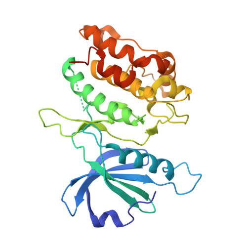
    6AAR

  • PubMed Abstract: 

    Death-associated protein kinase 1 (DAPK1) is a serine/threonine protein kinase that regulates apoptosis and autophagy. DAPK1 is considered to be a therapeutic target for amyloid-β deposition, endometrial adenocarcinomas and acute ischemic stroke. Here, the potent inhibitory activity of the natural anthraquinone purpurin against DAPK1 phosphorylation is shown. Thermodynamic analysis revealed that while the binding affinity of purpurin is similar to that of CPR005231, which is a DAPK1 inhibitor with an imidazopyridazine moiety, the binding of purpurin was more enthalpically favorable. In addition, the inhibition potencies were correlated with the enthalpic changes but not with the binding affinities. Crystallographic analysis of the DAPK1-purpurin complex revealed that the formation of a hydrogen-bond network is likely to contribute to the favorable enthalpic changes and that stabilization of the glycine-rich loop may cause less favorable entropic changes. The present findings indicate that purpurin may be a good lead compound for the discovery of inhibitors of DAPK1, and the observation of enthalpic changes could provide important clues for drug development.


  • Organizational Affiliation
    • Faculty of Pharmaceutical Sciences, University of Toyama, 2630 Sugitani, Toyama 930-0914, Japan.

Macromolecule Content 

  • Total Structure Weight: 34.05 kDa 
  • Atom Count: 2,392 
  • Modeled Residue Count: 269 
  • Deposited Residue Count: 293 
  • Unique protein chains: 1

Macromolecules

Find similar proteins by:|  3D Structure
Entity ID: 1
MoleculeChains  Sequence LengthOrganismDetailsImage
Death-associated protein kinase 1293Homo sapiensMutation(s): 0 
Gene Names: DAPK1DAPK
EC: 2.7.11.1
UniProt & NIH Common Fund Data Resources
Find proteins for P53355 (Homo sapiens)
Explore P53355 
Go to UniProtKB:  P53355
PHAROS:  P53355
GTEx:  ENSG00000196730 
Entity Groups
Sequence Clusters30% Identity50% Identity70% Identity90% Identity95% Identity100% Identity
UniProt GroupP53355
Sequence Annotations
Expand
Reference Sequence

Small Molecules

Ligands 1 Unique
IDChains Name / Formula / InChI Key2D Diagram3D Interactions
9TF
(Subject of Investigation/LOI)

Query on 9TF


Download:Ideal Coordinates CCD File
B [auth A]Purpurin
C14 H8 O5
BBNQQADTFFCFGB-UHFFFAOYSA-N
Binding Affinity Annotations 
IDSourceBinding Affinity
9TF BindingDB:  6AAR IC50: 890 (nM) from 1 assay(s)

Experimental Data & Validation

Experimental Data

  • Method: X-RAY DIFFRACTION
  • Resolution: 1.95 Å
  • R-Value Free:  0.220 (Depositor), 0.221 (DCC) 
  • R-Value Work:  0.180 (Depositor), 0.181 (DCC) 
  • R-Value Observed: 0.182 (Depositor) 
Space Group: P 21 21 21
Unit Cell:
Length ( Å )Angle ( ˚ )
a = 46.723α = 90
b = 62.892β = 90
c = 88.646γ = 90
Software Package:
Software NamePurpose
PHENIXrefinement
XDSdata reduction
XSCALEdata scaling
PHENIXphasing

Structure Validation

View Full Validation Report


Ligand Structure Quality Assessment 


Entry History 

Deposition Data

Revision History  (Full details and data files)

  • Version 1.0: 2019-07-24
    Type: Initial release
  • Version 1.1: 2020-05-13
    Changes: Database references
  • Version 1.2: 2024-03-27
    Changes: Data collection, Database references