Evolutionary conservation of protein dynamics: insights from all-atom molecular dynamics simulations of 'peptidase' domain of Spt16.
Gaur, N.K., Ghosh, B., Goyal, V.D., Kulkarni, K., Makde, R.D.(2021) J Biomol Struct Dyn : 1-13
- PubMed: 34971347 Search on PubMed
- DOI: https://doi.org/10.1080/07391102.2021.2021990
- Primary Citation Related Structures:
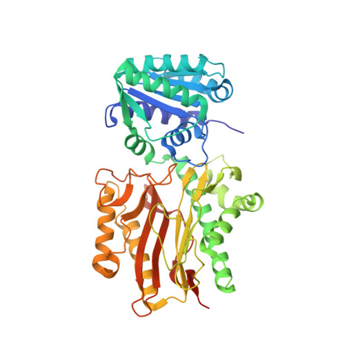
6A8M - PubMed Abstract:
Protein function is encoded in its sequence, manifested in its three-dimensional structure, and facilitated by its dynamics. Studies have suggested that protein structures with higher sequence similarity could have more similar patterns of dynamics. However, such studies of protein dynamics within and across protein families typically rely on coarse-grained models, or approximate metrics like crystallographic B-factors. This study uses µs scale molecular dynamics (MD) simulations to explore the conservation of dynamics among homologs of ∼50 kDa N-terminal module of Spt16 (Spt16N). Spt16N from Saccharomyces cerevisiae (Sc-Spt16N) and three of its homologs with 30-40% sequence identities were available in the PDB. To make our data-set more comprehensive, the crystal structure of an additional homolog (62% sequence identity with Sc-Spt16N) was solved at 1.7 Å resolution. Cumulative MD simulations of 6 µs were carried out on these Spt16N structures and on two additional protein structures with varying degrees of similarity to it. The simulations revealed that correlation in patterns of backbone fluctuations vary linearly with sequence identity. This trend could not be inferred using crystallographic B-factors. Further, normal mode analysis suggested a similar pattern of inter-domain (inter-lobe) motions not only among Spt16N homologs, but also in the M24 peptidase structure. On the other hand, MD simulation results highlighted conserved motions that were found unique for Spt16N protein, this along with electrostatics trends shed light on functional aspects of Spt16N.Communicated by Ramaswamy H. Sarma.
- Beamline Development and Application Section, Bhabha Atomic Research Centre, Mumbai, India.
Organizational Affiliation: