6A20 | pdb_00006a20

Crystal Structure of auto-inhibited Kinesin-3 KIF13B


Experimental Data Snapshot

  • Method: X-RAY DIFFRACTION
  • Resolution: 2.40 Å
  • R-Value Free: 
    0.249 (Depositor), 0.254 (DCC) 
  • R-Value Work: 
    0.193 (Depositor), 0.201 (DCC) 
  • R-Value Observed: 
    0.196 (Depositor) 

Starting Model: experimental
View more details

wwPDB Validation 3D Report Full Report

Validation slider image for 6A20

Ligand Structure Quality Assessment 


This is version 1.2 of the entry. See complete history

Literature

Coiled-coil 1-mediated fastening of the neck and motor domains for kinesin-3 autoinhibition.

Ren, J.Q.Wang, S.Chen, H.Wang, W.J.Huo, L.Feng, W.

(2018) Proc Natl Acad Sci U S A 115: E11933-E11942

  • DOI: https://doi.org/10.1073/pnas.1811209115
  • Primary Citation Related Structures: 
    6A1Z, 6A20

  • PubMed Abstract: 

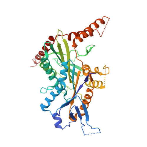
    In kinesin-3, the coiled-coil 1 (CC1) can sequester the preceding neck coil (NC) for autoinhibition, but the underlying mechanism is poorly understood. Here, we determined the structures of the uninhibited motor domain (MD)-NC dimer and inhibited MD-NC-CC1 monomer of kinesin-3 KIF13B. In the MD-NC-CC1 monomer, CC1 is broken into two short helices that unexpectedly interact with both the NC and the MD. Compared with the MD-NC dimer, the CC1-mediated integration of NC and MD not only blocks the NC dimer formation, but also prevents the neck linker (NL) undocking and the ADP release from the MD. Mutations of the essential residues in the interdomain interaction interface in the MD-NC-CC1 monomer restored the MD activity. Thus, CC1 fastens the neck domain and MD and inhibits both NC and NL. This CC1-mediated lockdown of the entire neck domain may represent a paradigm for kinesin autoinhibition that could be applicable to other kinesin-3 motors.


  • Organizational Affiliation
    • National Laboratory of Biomacromolecules, CAS Center for Excellence in Biomacromolecules, Institute of Biophysics, Chinese Academy of Sciences, 100101 Beijing, China.

Macromolecule Content 

  • Total Structure Weight: 49.73 kDa 
  • Atom Count: 3,297 
  • Modeled Residue Count: 395 
  • Deposited Residue Count: 437 
  • Unique protein chains: 1

Macromolecules

Find similar proteins by:|  3D Structure
Entity ID: 1
MoleculeChains  Sequence LengthOrganismDetailsImage
Kinesin family member 13B437Rattus norvegicusMutation(s): 1 
Gene Names: Kif13b
UniProt
Find proteins for A0A0G2K8Z9 (Rattus norvegicus)
Explore A0A0G2K8Z9 
Go to UniProtKB:  A0A0G2K8Z9
Entity Groups
Sequence Clusters30% Identity50% Identity70% Identity90% Identity95% Identity100% Identity
UniProt GroupA0A0G2K8Z9
Sequence Annotations
Expand
Reference Sequence

Experimental Data & Validation

Experimental Data

  • Method: X-RAY DIFFRACTION
  • Resolution: 2.40 Å
  • R-Value Free:  0.249 (Depositor), 0.254 (DCC) 
  • R-Value Work:  0.193 (Depositor), 0.201 (DCC) 
  • R-Value Observed: 0.196 (Depositor) 
Space Group: P 32
Unit Cell:
Length ( Å )Angle ( ˚ )
a = 75.07α = 90
b = 75.07β = 90
c = 91.71γ = 120
Software Package:
Software NamePurpose
PHENIXrefinement
HKL-3000data reduction
HKL-3000data scaling
PHASERphasing

Structure Validation

View Full Validation Report


Ligand Structure Quality Assessment 


Entry History 

Deposition Data

Revision History  (Full details and data files)

  • Version 1.0: 2018-11-21
    Type: Initial release
  • Version 1.1: 2019-01-02
    Changes: Data collection, Database references
  • Version 1.2: 2023-11-22
    Changes: Data collection, Database references, Derived calculations, Refinement description