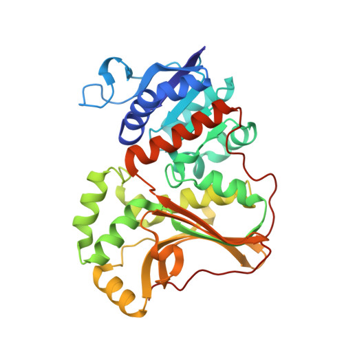
6A0T

Homoserine dehydrogenase K99A mutant from Thermus thermophilus HB8 complexed with HSE and NADP+


Experimental Data Snapshot

  • Method: X-RAY DIFFRACTION
  • Resolution: 1.87 Å
  • R-Value Free: 0.167 
  • R-Value Work: 0.137 
  • R-Value Observed: 0.138 

Starting Model: experimental
View more details

wwPDB Validation   3D Report Full Report


Ligand Structure Quality Assessment 


This is version 2.1 of the entry. See complete history


Literature

The crystal structure of homoserine dehydrogenase complexed with l-homoserine and NADPH in a closed form

Akai, S.Ikushiro, H.Sawai, T.Yano, T.Kamiya, N.Miyahara, I.

(2019) J Biochem 165: 185-195

  • DOI: https://doi.org/10.1093/jb/mvy094
  • Primary Citation of Related Structures:  
    6A0R, 6A0S, 6A0T, 6A0U

  • PubMed Abstract: 

    Homoserine dehydrogenase from Thermus thermophilus (TtHSD) is a key enzyme in the aspartate pathway that catalyses the reversible conversion of l-aspartate-β-semialdehyde to l-homoserine (l-Hse) with NAD(P)H. We determined the crystal structures of unliganded TtHSD, TtHSD complexed with l-Hse and NADPH, and Lys99Ala and Lys195Ala mutant TtHSDs, which have no enzymatic activity, complexed with l-Hse and NADP+ at 1.83, 2.00, 1.87 and 1.93 Å resolutions, respectively. Binding of l-Hse and NADPH induced the conformational changes of TtHSD from an open to a closed form: the mobile loop containing Glu180 approached to fix l-Hse and NADPH, and both Lys99 and Lys195 could make hydrogen bonds with the hydroxy group of l-Hse. The ternary complex of TtHSDs in the closed form mimicked a Michaelis complex better than the previously reported open form structures from other species. In the crystal structure of Lys99Ala TtHSD, the productive geometry of the ternary complex was almost preserved with one new water molecule taking over the hydrogen bonds associated with Lys99, while the positions of Lys195 and l-Hse were significantly retained with those of the wild-type enzyme. These results propose new possibilities that Lys99 is the acid-base catalytic residue of HSDs.


  • Organizational Affiliation

    Graduate School of Science, Osaka City University, Osaka, Japan.


Macromolecules
Find similar proteins by:  (by identity cutoff)  |  3D Structure
Entity ID: 1
MoleculeChains Sequence LengthOrganismDetailsImage
Homoserine dehydrogenaseA [auth B],
B [auth A]
332Thermus thermophilus HB8Mutation(s): 0 
Gene Names: TTHA0489
EC: 1.1.1.3
UniProt
Find proteins for Q5SL04 (Thermus thermophilus (strain ATCC 27634 / DSM 579 / HB8))
Explore Q5SL04 
Go to UniProtKB:  Q5SL04
Entity Groups  
Sequence Clusters30% Identity50% Identity70% Identity90% Identity95% Identity100% Identity
UniProt GroupQ5SL04
Sequence Annotations
Expand
  • Reference Sequence
Small Molecules
Ligands 6 Unique
IDChains Name / Formula / InChI Key2D Diagram3D Interactions
NAP
Query on NAP

Download Ideal Coordinates CCD File 
E [auth B],
O [auth A]
NADP NICOTINAMIDE-ADENINE-DINUCLEOTIDE PHOSPHATE
C21 H28 N7 O17 P3
XJLXINKUBYWONI-NNYOXOHSSA-N
CXS
Query on CXS

Download Ideal Coordinates CCD File 
F [auth B]3-CYCLOHEXYL-1-PROPYLSULFONIC ACID
C9 H19 N O3 S
PJWWRFATQTVXHA-UHFFFAOYSA-N
HSE
Query on HSE

Download Ideal Coordinates CCD File 
D [auth B],
N [auth A]
L-HOMOSERINE
C4 H9 N O3
UKAUYVFTDYCKQA-VKHMYHEASA-N
GOL
Query on GOL

Download Ideal Coordinates CCD File 
K [auth B],
L [auth B],
V [auth A],
W [auth A],
X [auth A]
GLYCEROL
C3 H8 O3
PEDCQBHIVMGVHV-UHFFFAOYSA-N
FMT
Query on FMT

Download Ideal Coordinates CCD File 
G [auth B]
H [auth B]
I [auth B]
J [auth B]
P [auth A]
G [auth B],
H [auth B],
I [auth B],
J [auth B],
P [auth A],
Q [auth A],
R [auth A],
S [auth A],
T [auth A],
U [auth A]
FORMIC ACID
C H2 O2
BDAGIHXWWSANSR-UHFFFAOYSA-N
NA
Query on NA

Download Ideal Coordinates CCD File 
C [auth B],
M [auth A]
SODIUM ION
Na
FKNQFGJONOIPTF-UHFFFAOYSA-N
Experimental Data & Validation

Experimental Data

  • Method: X-RAY DIFFRACTION
  • Resolution: 1.87 Å
  • R-Value Free: 0.167 
  • R-Value Work: 0.137 
  • R-Value Observed: 0.138 
  • Space Group: P 32 2 1
Unit Cell:
Length ( Å )Angle ( ˚ )
a = 121.401α = 90
b = 121.401β = 90
c = 145.089γ = 120
Software Package:
Software NamePurpose
REFMACrefinement
HKL-2000data reduction
HKL-2000data scaling
MOLREPphasing

Structure Validation

View Full Validation Report


Ligand Structure Quality Assessment 


Entry History 

Deposition Data

Revision History  (Full details and data files)

  • Version 1.0: 2018-11-28
    Type: Initial release
  • Version 1.1: 2019-01-30
    Changes: Data collection, Database references
  • Version 2.0: 2023-11-15
    Changes: Atomic model, Data collection, Database references
  • Version 2.1: 2023-11-22
    Changes: Refinement description